Membranoproliferative Glomerulonephritis with Striated Ultrastructural Deposits with Significantly Elevated Fibrinogen and Fibronectin on Mass Spectrometry Analysis: A Case Report and Literature Review
- PMID: 39947166
- PMCID: PMC12215155
- DOI: 10.1159/000544709
Membranoproliferative Glomerulonephritis with Striated Ultrastructural Deposits with Significantly Elevated Fibrinogen and Fibronectin on Mass Spectrometry Analysis: A Case Report and Literature Review
Abstract
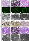
Glomerular diseases with organized deposits can be classified into various etiologies. A diagnostic algorithm based on clinical and pathological findings has been proposed. However, some cases cannot be diagnosed using existing algorithms. Here, we report the case of a 77-year-old man diagnosed with membranoproliferative glomerulonephritis (MPGN) with striated ultrastructural deposits, micro-filament-like substructures with straight bands arranged in parallel in the subendothelial space by two sequential renal biopsies. His examinations and clinical findings were incompatible with known glomerular diseases with organized deposits. Dialysis was initiated 10 months after the second biopsy procedure. Furthermore, we report the first mass spectrometry analysis of laser micro-dissected glomeruli with striated ultrastructural deposits, which revealed significant levels of fibrinogen and fibronectin. Immunostaining was positive for fibrinogen, fibrin, and fibronectin in the subendothelial space. These findings suggest that the deposits were composed of a fibrin-fibronectin complex and that accumulation of these fibrin-fibronectin complexes possibly induced endothelial injury, leading to MPGN. We also reviewed the literature on the clinical and pathological characteristics of the four cases with striated ultrastructural deposits. Our investigation showed that all patients had the MPGN pattern and striated ultrastructural deposits in the subendothelial space, and all underwent hemodialysis within 3 years of renal biopsy. Clinicians should be aware of the findings of glomerulonephritis with striated ultrastructural deposits since this disease may be a new entity and has a poor prognosis.
Keywords: Deposit; Fibrin; Fibrinogen; Fibronectin; Mass spectrometry; Membranoproliferative glomerulonephritis.
© 2025 The Author(s). Published by S. Karger AG, Basel.
Conflict of interest statement
The authors have no conflicts of interest to declare.
Figures


References
-
- Herrera GA, Turbat-Herrera EA. Renal diseases with organized deposits: an algorithmic approach to classification and clinicopathologic diagnosis. Arch Pathol Lab Med. 2010;134(4):512–31. - PubMed
-
- Alan SLY, Chertow GM, Luyckx VA, Marsden PA, Skorecki K, Taal MW, et al. Brenner & Rector’s THE KIDNEY eleventh edition, [chapter 31]. p. 1069.
-
- Vrana JA, Gamez JD, Madden BJ, Theis JD, Bergen HR 3rd, Dogan A. Classification of amyloidosis by laser microdissection and mass spectrometry-based proteomic analysis in clinical biopsy specimens. Blood. 2009;114(24):4957–9. - PubMed
-
- Gonzalez Suarez ML, Zhang P, Nasr SH, Sathick IJ, Kittanamongkolchai W, Kurtin PJ, et al. The sensitivity and specificity of the routine kidney biopsy immunofluorescence panel are inferior to diagnosing renal immunoglobulin-derived amyloidosis by mass spectrometry. Kidney Int. 2019;96(4):1005–9. - PubMed

