. 2025 Jun 5;65(6):2400276.
doi: 10.1183/13993003.00276-2024. Print 2025 Jun.

Association of fibrotic-related extracellular vesicle microRNAs with lung involvement in systemic sclerosis

Affiliations

Association of fibrotic-related extracellular vesicle microRNAs with lung involvement in systemic sclerosis

Julien Guiot et al. Eur Respir J. .

Abstract

Background: There is a pressing need to identify early biomarkers of lung involvement in systemic sclerosis to start antifibrotic therapy as soon as possible. We aimed to identify extracellular vesicle-derived microRNAs (miRNAs) that are differentially expressed between systemic sclerosis patients with and without interstitial lung disease, and to explore their diagnostic value and functional properties.

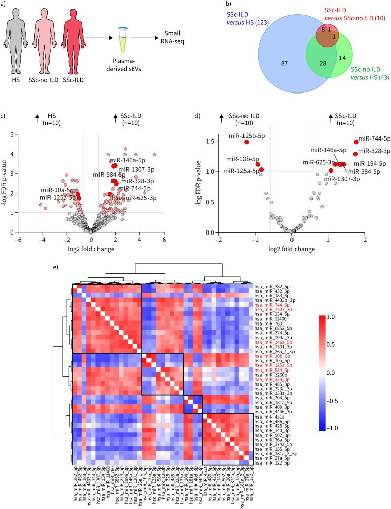
Methods: Small extracellular vesicles derived from plasma were isolated from 91 well-characterised patients with systemic sclerosis with (n=45) and without (n=46) interstitial lung disease and 43 matched healthy subjects. Small RNA-sequencing followed by quantitative reverse transcriptase PCR were used to identify and validate small extracellular vesicle miRNAs associated with systemic sclerosis-associated interstitial lung disease. Correlations between systemic sclerosis-associated interstitial lung disease miRNAs and clinical parameters were assessed, as well as the effect of related miRNAs/small extracellular vesicles on fibrosis.

Results: We identified a four-miRNA signature associated with interstitial lung disease in the context of systemic sclerosis (miR-584-5p, miR-744-5p, miR-1307-3p and miR-10b-5p) (area under the receiver operating characteristic curve 0.85, 95% CI 0.76-0.94; p<0.0001). Deeper analysis revealed a correlation of these candidates with pulmonary function tests (diffusing capacity of the lung for carbon monoxide and forced vital capacity), highlighting their use in monitoring lung fibrosis progression in systemic sclerosis patients. Furthermore, small extracellular vesicle miRNAs associated with systemic sclerosis-associated interstitial lung disease are positively correlated with and enriched in circulating lymphocytes, suggesting that these immune cells are their cellular source. Finally, functional studies highlighted altered functional properties of small extracellular vesicles in the context of systemic sclerosis-associated interstitial lung disease, mainly due to the transfer of profibrotic miR-584-5p in lung fibroblasts.

Conclusions: Our small extracellular vesicle-based biomarker approach identified a promising four‑miRNA signature characteristic of interstitial lung disease in systemic sclerosis patients. Furthermore, the profibrotic properties of small extracellular vesicles associated with systemic sclerosis-associated interstitial lung disease suggest a prominent role of these vesicles in systemic sclerosis severity.


Conflict of interest statement

Conflict of interest: R. Louis reports grants from Chiesi, AstraZeneca and GSK; consultancy fees from AstraZeneca and GSK; payment or honoraria for lectures, presentations, manuscript writing or educational events from AstraZeneca and GSK; and participation on a data safety monitoring board or advisory board with AstraZeneca. J. Guiot reports payment or honoraria for lectures, presentations, manuscript writing or educational events from Boehringer Ingelheim, Janssens, GSK, Roche and Chiesi; support for attending meetings from Chiesi, Roche, Janssens, Boehringer Ingelheim and AstraZeneca; patents planned, issued or pending with Radiomics (Oncoradiomics SA); and participation on a data safety monitoring board or advisory board with GSK, Janssens, Chiesi, AstraZeneca and MSD. The rest of the authors declare that they have no conflicts of interest.

Figures

None
Overview of the study. SSc: systemic sclerosis; ILD: interstitial lung disease; HS: healthy subjects; sEV: small extracellular vesicle; RT-qPCR: quantitative reverse transcriptase PCR; FC: fold change; ECM: extracellular matrix.
FIGURE 1
FIGURE 1
Workflow of study design and characterisation of plasma small extracellular vesicle (sEVs). a) Schematic representation of the approach used to identify a microRNA (miRNA) signature for systemic sclerosis (SSc) with interstitial lung disease (ILD). b) Particle size distribution analysis of sEVs isolated from plasma of SSc patients (with/without ILD) and healthy control subjects (HS). c) Quantification of the mean particle size and the particle concentration (particles·mL−1) of indicated EVs (n=2 biological replicates). d) Western blot analysis of exosomal markers (CD9, CD81, CD63, syntenin) and mitochondrial cytochrome c in protein extracts from plasma sEVs of SSc patients (with/without ILD) and HS and from cell lysates. 5 μg of sEV or cell protein were loaded in the Western blot for every sample. RT-qPCR: quantitative reverse transcriptase PCR; ROC: receiver operating characteristic.
FIGURE 2
FIGURE 2
Identification of small extracellular vesicle (sEV)-related microRNAs (miRNAs) specific to lung involvement in systemic sclerosis (SSc) patients via small RNA-sequencing (RNA-seq). a) Overview of the procedure to identify sEV miRNAs associated with SSc-associated interstitial lung disease (ILD). Small RNA-seq of plasma sEVs from SSc patients with ILD (SSc-ILD, n=10), without ILD (SSc-no ILD, n=10) and healthy control subjects (HS, n=10). b) Venn diagram illustrating the number of shared and distinct miRNAs across the differential expression analysis: SSc-ILD versus HS, SSc-no ILD versus HS and SSc-ILD versus SSc-no ILD (fold change >1.5 and false discovery rate (FDR) <0.1). c) Volcano plot showing plasma sEV miRNAs that were differentially expressed between HS (n=10) and SSc-ILD patients (n=10). There were 123 miRNAs differentially expressed in SSc-ILD patients compared to HS (fold change >1.5 and FDR<0.1): 80 upregulated and 43 downregulated. Highlighted are miRNAs that were associated with ILD in SSc patients. d) Volcano plot showing plasma sEV miRNAs that were differentially expressed between SSc-ILD patients (n=10) and SSc-no ILD patients (n=10). There were 10 miRNAs differentially expressed in SSc-ILD patients compared to SSc-no ILD patients (fold change >1.5 and FDR<0.1): seven upregulated and three downregulated. e) Clusters and correlations of sEV miRNAs measured by small RNA-seq in SSc-ILD patients. The heatmap represents a hierarchical cluster analysis conducted upon a Spearman correlation network of miRNA levels in SSc patients that were found to be differentially expressed between SSc-ILD patients and HS (fold change >1.5, FDR<0.02) and for which a critical role in lung fibrosis and inflammation has been shown previously.
FIGURE 3
FIGURE 3
Quantitative reverse transcriptase PCR (RT-qPCR) validation of microRNAs (miRNAs) specific to lung involvement in systemic sclerosis (SSc) patients and assessment of their diagnostic value. a) RT-qPCR of specific miRNAs miR-584-5p, miR-744-5p, miR-1307-3p and miR-10b-5p in plasma small extracellular vesicles (sEVs) of SSc patients with interstitial lung disease (ILD) (SSc-ILD, n=35), without ILD (SSc-no ILD, n=36) and healthy control subjects (HS, n=33). Data were non-normally distributed and analysed using a non-parametric Kruskal–Wallis test, followed by Benjamini, Krieger and Yekutieli's false discovery rate (FDR) correction for multiple comparisons. FC: fold change. b) Receiver operator characteristic (ROC) curves with corresponding area under the curves (AUCs) for comparing the ability of the four-miRNA signature (miR-584-5p+miR-744-5p+miR-1307-3p+miR-10b-5p) and diffusing coefficient of the lungs for carbon monoxide (DLCO) (%) to discriminate SSc-ILD patients (n=35) from SSc-no ILD patients (n=36). c) Molecular Signatures Database (MSigDB) hallmark targeted by SSc-ILD-related miRNAs (miR-584-5p, miR-744-5p, miR-1307-3p and miR-10b-5p). In silico analysis was performed with the DIANA-miRPath v4.0 tool using Tarbase v.8. TGF-β: transforming growth factor β. *: FDR<0.05; **: FDR<0.01; ***: FDR<0.001; ****: FDR<0.0001.
FIGURE 4
FIGURE 4
Correlation analysis of small extracellular vesicle (sEV)-related microRNAs (miRNAs) and lung function in systemic sclerosis (SSc) patients. a, e, i) Heatmaps showing correlations between lung function (assessed by forced vital capacity (FVC) and diffusing capacity of the lung for carbon monoxide (DLCO)) and the levels of SSc-associated interstitial lung disease (ILD)-related miRNAs (miR-584-5p, miR-744-5p, miR-1307‑3p and miR-10b-5p) in a) all SSc patients, e) SSc patients without ILD (SSc-no ILD) and i) SSc patients with ILD (SSc-ILD). b, f, j) Correlation between FVC and the levels of sEV miR-584-5p in b) all SSc patients, f) SSc-no ILD patients and j) SSc-ILD patients. c, g, k) Correlation between FVC and the levels of sEV miR-1307-3p in c) all SSc patients, g) SSc-no ILD patients and k) SSc-ILD patients. d, h, l) Correlation between DLCO and the levels of sEV miR-10b-5p in d) all SSc patients, h) SSc-no ILD patients and l) SSc-ILD patients. Data were non-normally distributed and analysed using Spearman correlation (two-tailed). *: p<0.05; **: p<0.01.
FIGURE 5
FIGURE 5
Cellular origin of small extracellular vesicle (sEV) microRNAs (miRNAs) associated with lung involvement in systemic sclerosis (SSc) patients. a, b) Heatmaps showing correlations of SSc-associated interstitial lung disease (ILD)-related miRNAs (miR-584-5p, miR-744-5p, miR-1307‑3p and miR-10b-5p) with plasma-derived cells (neutrophils, lymphocytes and monocytes) in a) SSc patients without ILD (SSc-no ILD) and b) SSc‑ILD patients. Data were normally distributed and analysed using Pearson correlation (two-tailed). c–e) Correlation between plasma levels of sEV miR-744-5p and c) circulating neutrophils, d) lymphocytes and e) monocytes in SSc-ILD patients. f) Schematic representation of the purification of circulating neutrophils and lymphocytes from buffy coats of healthy control subjects (HS). g) Purity was evaluated by flow cytometry after staining of lymphocytic fraction and polymorphonuclear leukocyte (PMN) fraction with anti-CD3 allophyocyanin (APC) and anti-CD19 Brilliant Violet 421 (BV421). h) Expression level of miR-584-5p and miR-744-5p in circulating neutrophils and lymphocytes purified from buffy coats of HS. Data were analysed using ordinary two-way ANOVA with Sidak's correction. PBMC: peripheral blood mononuclear cell. *: p<0.05.
FIGURE 6
FIGURE 6
Plasma small extracellular vesicle (sEVs) from patients with systemic sclerosis (SSc) with interstitial lung disease (ILD) are able to transfer miR-584-5p and miR-744-5p to recipient lung fibroblasts. a) Schematic representation of the treatment of lung fibroblast cell line MRC-5 with plasma sEVs (5 µg·mL−1) derived from 1) healthy control subjects (HS), 2) SSc patients without ILD (SSc-no ILD) or 3) SSc-ILD patients for 24 h. b) Transcript levels of SSc-ILD-related microRNAs (miRNAs) (miR-584-5p and miR-744-5p) in lung fibroblast cell line MRC-5 treated with corresponding sEVs (5 µg·mL−1) for 24 h assessed by quantitative reverse transcriptase PCR (HS, n=12; SSc-no ILD, n=10; SSc-ILD, n=8). Data are expressed as median±IQR and were analysed using ordinary one-way ANOVA with Tukey's correction (normally distributed data). c) Kinetics of the expression of mature miR-584-5p and miR-744-5p and their precursors (pre-miR-584 and pre-miR-744, respectively) after treatment of MRC-5 cells with SSc-ILD sEVs (5 μg·mL−1) (n=3). *: p<0.05.
FIGURE 7
FIGURE 7
Plasma small extracellular vesicle (sEVs) from patients with systemic sclerosis (SSc) with interstitial lung disease (ILD) have miR-584-5p-dependent profibrotic properties. a) miR-584-5p overexpression increases the expression of profibrotic genes (fibronectin (FN), actin α2 smooth muscle (ACTA2), collagen type I α1 (COL1A1) and collagen type III α1 (COL3A1) in transforming growth factor β1 (TGF-β1)-treated MRC-5 cells (n=4), as assessed by quantitative reverse transcriptase PCR (qRT-PCR). b) miR-584-5p inhibition decreases the expression of profibrotic genes (FN and ACTA2) in TGF-β1-treated MRC-5 cells (n=3), as assessed by qRT-PCR. Data are expressed as median±IQR and were analysed using multiple paired t-test (a and b). c) Schematic representation of the treatment of lung fibroblasts (MRC-5) with plasma sEVs (5 µg·mL−1) derived from 1) healthy control subjects (HS), 2) SSc patients without ILD (SSc-no ILD) or 3) SSc‑ILD patients for 24 h, followed by TGF-β stimulation for 4 h. d) Plasma sEVs from SSc-ILD increased the expression of profibrotic genes (FN, ACTA2, COL1A1 and COL3A1) in MRC-5 cells compared to the ones from SSc-no ILD, as assessed by qRT-PCR (HS, n=12; SSc-no ILD, n=9; SSc-ILD, n=13). Data are expressed as median±IQR and were analysed using ordinary one-way ANOVA with Tukey's correction (for normally distributed data) or Kruskal–Wallis test with Dunn's correction (for non-normally distributed data). e) miR-584-5p overexpression increases the protein expression of FN in TGF-β1-treated MRC-5 cells (n=3), as assessed by flow cytometry. f) Plasma sEVs from SSc-ILD increase the protein expression of FN in MRC-5 cells compared to those from SSc-no ILD, as assessed by Western blot. g) Inhibiting miR-584-5p in MRC-5 cells restores the sEV-dependent regulation of profibrotic genes in treated cells. Data are expressed relative to cells without sEV treatment (n=3). Data are expressed as mean±sd and were analysed using multiple unpaired t-test. All statistical analyses were two-tailed. ctrl: control. *: p<0.05; **: p<0.01; ***: p<0.001.

Comment in

References

    1. Nihtyanova SI, Denton CP. Pathogenesis of systemic sclerosis associated interstitial lung disease. J Scleroderma Relat Disord 2020; 5: 6–16. doi:10.1177/2397198320903867 - DOI - PMC - PubMed
    1. Petelytska L, Bonomi F, Cannistrà C, et al. . Heterogeneity of determining disease severity, clinical course and outcomes in systemic sclerosis-associated interstitial lung disease: a systematic literature review. RMD Open 2023; 9: e003426. doi:10.1136/rmdopen-2023-003426 - DOI - PMC - PubMed
    1. Khanna D, Tashkin DP, Denton CP, et al. . Etiology, risk factors, and biomarkers in systemic sclerosis with interstitial lung disease. Am J Respir Crit Care Med 2020; 201: 650–660. doi:10.1164/rccm.201903-0563CI - DOI - PMC - PubMed
    1. Hoffmann-Vold A-M, Fretheim H, Halse A-K, et al. . Tracking impact of interstitial lung disease in systemic sclerosis in a complete nationwide cohort. Am J Respir Crit Care Med 2019; 200: 1258–1266. doi:10.1164/rccm.201903-0486OC - DOI - PubMed
    1. Perelas A, Silver RM, Arrossi AV, et al. . Systemic sclerosis-associated interstitial lung disease. Lancet Respir Med 2020; 8: 304–320. doi:10.1016/S2213-2600(19)30480-1 - DOI - PubMed