. 2025 Feb 13;16(1):1604.
doi: 10.1038/s41467-025-56902-x.

Design of quinoline SARS-CoV-2 papain-like protease inhibitors as oral antiviral drug candidates

Affiliations

Design of quinoline SARS-CoV-2 papain-like protease inhibitors as oral antiviral drug candidates

Prakash Jadhav et al. Nat Commun. .

Abstract

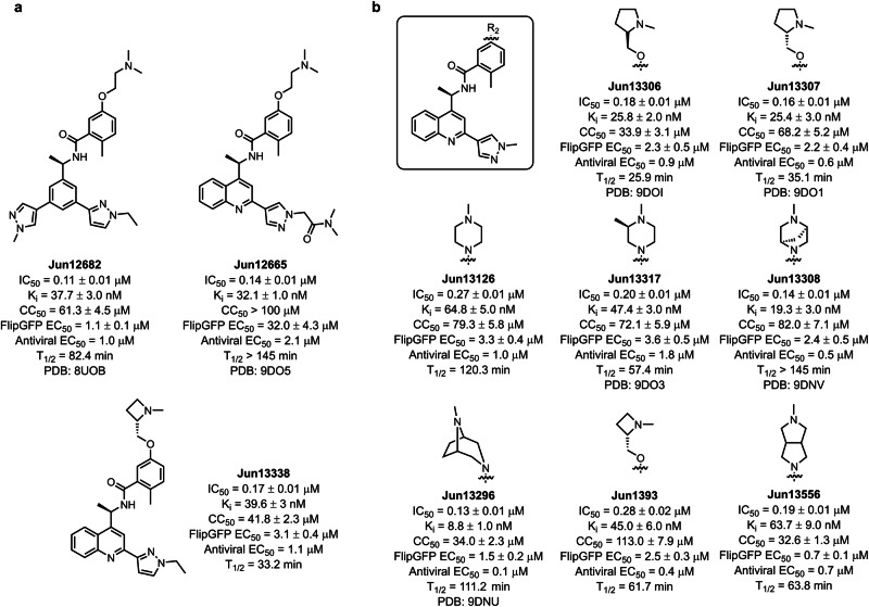
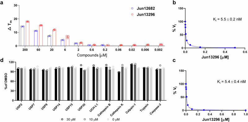
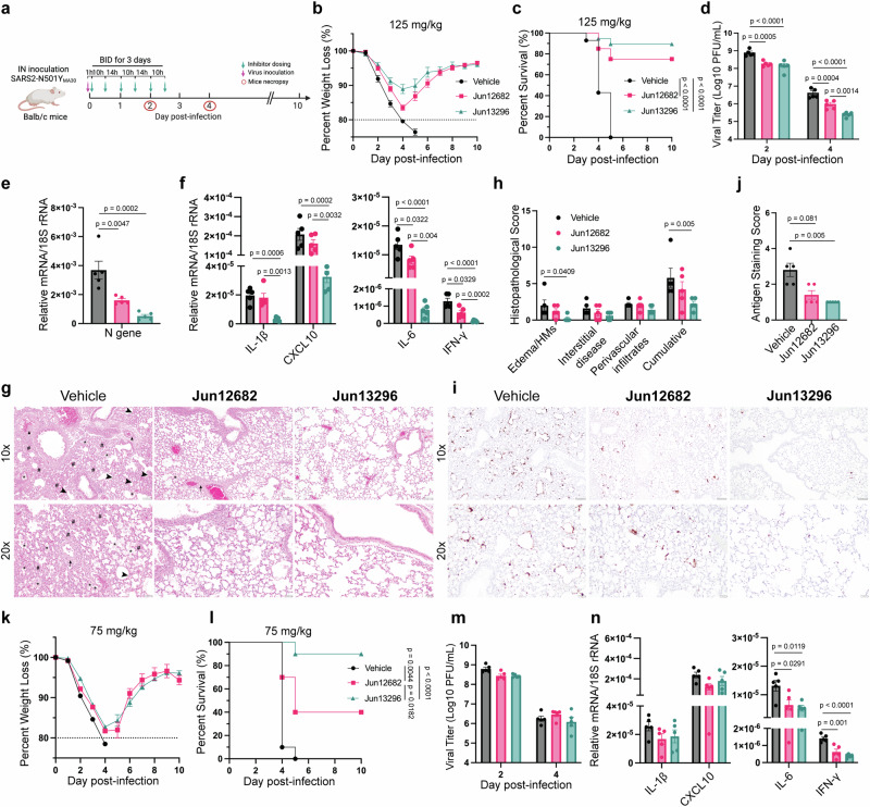
The ever-evolving SARS-CoV-2 variants necessitate the development of additional oral antivirals. This study presents the systematic design of quinoline-containing SARS-CoV-2 papain-like protease (PLpro) inhibitors as potential oral antiviral drug candidates. By leveraging the recently discovered Val70Ub binding site in PLpro, we designed a series of quinoline analogs demonstrating potent PLpro inhibition and antiviral activity. Notably, the X-ray crystal structures of 6 lead compounds reveal that the 2-aryl substitution can occupy either the Val70Ub site as expected or the BL2 groove in a flipped orientation. The in vivo lead Jun13296 exhibits favorable pharmacokinetic properties and potent inhibition against SARS-CoV-2 variants and nirmatrelvir-resistant mutants. In a mouse model of SARS-CoV-2 infection, oral treatment with Jun13296 significantly improves survival, reduces body weight loss and lung viral titers, and prevents lung tissue damage. These results underscore the potential of quinoline PLpro inhibitors as promising oral SARS-CoV-2 antiviral candidates, instilling hope for the future of SARS-CoV-2 treatment.


Conflict of interest statement

Competing interests: Rutgers, the State University of New Jersey, has applied for a patent US2024382494A1 covering the PLpro inhibitors reported in this study and related compounds, which has been published and is pending. J.W. is listed as an inventor. The remaining authors declare no competing interests.

Figures

Fig. 1
Fig. 1. Design of quinoline SARS-CoV-2 PLpro inhibitors.
a Superposition of the X-ray crystal structures of SARS-CoV-2 PLpro with Jun11313 (green) (PDB: 8UVM) and ubiquitin (orange) (PDB: 6XAA). b Superposition of the X-ray crystal structures of SARS-CoV-2 PLpro with Jun11313 (green) (PDB: 8UVM) and GRL0617 (yellow) (PDB: 7JRN). c Design of the quinoline PLpro inhibitors based on Jun11313 and GRL0617. Source data of (c) are provided as a Source Data file.
Fig. 2
Fig. 2. Representative quinoline SARS-CoV-2 PLpro inhibitors.
a Chemical structure and in vitro activities of Jun12682, Jun12665, and Jun13338. b Chemical structures and in vitro activities of quinoline analogs with diverse amine substituents. IC50, half maximal inhibitory concentration in the FRET enzymatic assay; Ki, inhibitory constant in the FRET enzymatic assay; CC50: half maximal toxicity concentration in Vero cells, CC50 values are mean ± S.D. of three technical repeats; EC50, half maximal effective concentration in the FlipGFP and antiviral assays, EC50 values are mean ± standard deviation of three technical repeats; T1/2, half-life in mouse liver microsomal stability assay. Source data are provided as a source data file.
Fig. 3
Fig. 3. In vitro and In vivo PK SARS-CoV-2 PLpro inhibitors.
a Plasma drug concentration of Jun13306, Jun13307, Jun13308, and Jun13317 in C57BL/6J mice (6 to 8 weeks old) following p.o. administration of 50 mg/kg of compounds in 0.5% methylcellulose and 2% Tween 80 in water (n = 3 per group). b Plasma drug concentrations of Jun13338, Jun1393, Jun13126, and Jun13296 in C57BL/6J mice (6 to 8 weeks old) following p.o. administration of 50 mg/kg of compounds in 0.5% methylcellulose and 2% Tween 80 in water (n = 3 per group). c Plasma drug concentrations of Jun13296 in C57BL/6J mice (6 to 8 weeks old) following p.o. administration of 50 mg/kg and i.v. injection of 10 mg/kg (n = 3 per group) of compound. The error bars are mean ± s.d. d In vivo pharmacokinetic parameters of Jun13296 in C57BL/6J mice. e In vitro pharmacokinetic parameters of Jun13296. T1/2, half-life; Tmax, time for maximal concentration; Cmax, maximum plasma concentration; AUC0-t, area under the curve from time zero to time t; AUCt-∞, area under the curve from time t to infinity; CL, clearance; MRT, mean residence time; Vss, volume of distribution; F, oral bioavailability. Source data are provided as a source data file.
Fig. 4
Fig. 4. Mechanistic studies of Jun13296.
a Differential scanning fluorimetry assay of Jun13296 in stabilizing SARS-CoV-2 PLpro. Jun12682 was included as a positive control for comparison. Data from Jun12682 is the mean of two repeats, and data from Jun13296 is the mean ± standard deviation of three technical repeats. b Ki plot of Jun13296 in inhibiting SARS-CoV-2 PLpro hydrolysis of ISG15-AMC. c Ki plot of Jun13296 in inhibiting SARS-CoV-2 PLpro hydrolysis of Ub-AMC. d Counter screening of Jun13296 against host proteases USP2, USP7, USP8, USP14, USP15, USP30, UCH-L1, cathepsin B, cathepsin K, calpain-1, trypsin, and caspase 3. Data in (d) are presented as mean ± standard deviation of two technical repeats. Source data are provided as a source data file.
Fig. 5
Fig. 5. X-ray crystal structures of SARS-CoV-2 PLpro with quinoline inhibitors.
Binding of quinoline inhibitors to SARS-CoV-2 PLpro. af Interactions of Jun12665 (orange), Jun13306 (yellow), Jun13307 (green), Jun13317 (light blue), Jun13308 (magenta), and Jun13296 (bright orange) with PLpro are conserved (residues within 5 Å of the inhibitor are shown as light brown sticks). Hydrogen bonds are indicated by black dashed lines, van der Waals contacts by red dashed lines, and π–π interactions by light green dashed lines. g The superposition of Jun12665 onto Jun13296 represents the flipped orientation of terminal amide carbonyl groups toward the BL2 groove. h Superposition of Jun13306 (yellow) and Jun13307 (green) containing the R and S pyrrolidine substituents both form similar electrostatic interactions with the Glu167 carboxylate.
Fig. 6
Fig. 6. Broad-spectrum antiviral activity of SARS-CoV-2 PLpro inhibitors.
The antiviral activity of nirmatrelvir, Jun12682, and Jun13296 was tested in the plaque assay using Vero-ACE2-TMPRSS2 (Vero-AT) cells. a Plaque assay EC50 plots of nirmatrelvir. b Plaque assay EC50 plots for Jun12682. c Plaque assay EC50 plots of Jun13296. EC50 values are mean ± standard deviation of two technical repeats. Source data are provided as a source data file.
Fig. 7
Fig. 7. In vivo antiviral efficacy of Jun13296.
a Experimental design for twice-a-day (BID) treatment for 3 days. b Mouse body weight loss and c survival rate of the mice receiving a 125 mg/kg BID_3 treatment. Data in (b, c) are pooled results of four independent experiments and are shown as mean ± standard error of the mean (SEM) (n = 14, 20, 19 mice for the Vehicle, Jun12682, and Jun13296 group, respectively). d Viral titers in lungs (n = 5 per group). Quantitative PCR analysis of viral nucleocapsid gene (e) and cellular cytokines (f) in lungs (n = 5 per group). g Haematoxylin and eosin (H&E) staining of lungs collected 4 DPI (n = 5 per group). Lungs exhibited airway edema (asterisks), hyaline membranes (HM, arrowheads), perivascular cuffing (arrows), and interstitial thickness (number sign). Scale bars, 100 mm (top) and 50 mm (bottom). h Quantification of the pathological lesions (g). i Immunostaining of lungs collected 4 DPI (n = 5 per group) for SARS-CoV-2 nucleocapsid protein (brown color staining). Scale bars, 100 µm (top) and 50 µm (bottom). j Summary scores of nucleocapsid immunostaining of lungs. k Mouse body weight loss and l survival rate of the mice receiving a 75 mg/kg BID_3 treatment. Data are pooled results of two independent experiments (n = 10 per group) and are shown as mean ± SEM. m Viral titers in lungs (n = 5 per group). n Quantitative PCR analysis of cellular cytokines in lungs collected 2 DPI (n = 5 per group). Data in (c, l) are shown as mean ± SEM, and the p values were determined using a log-rank (Mantel-Cox) test. Data in (d, m) are shown as mean ± SEM and analyzed with a two-way ANOVA Tukey’s multiple comparison test. Data in (e, f, n) are shown as mean ± SEM and analyzed with a one-way ANOVA multiple comparison test for each tested gene. Data in (h, j) are mean ± SEM and analyzed with the Krushal-Wallis multiple comparisons test for each category. Source data are provided as a source data file. a was created with Biorender.com. Li, K. (2025) https://BioRender.com/q16l908.

References

    1. Li, G., Hilgenfeld, R., Whitley, R. & De Clercq, E. Therapeutic strategies for COVID-19: progress and lessons learned. Nat. Rev. Drug Discov.22, 449–475 (2023). - PMC - PubMed
    1. Shoichet, B. K. & Craik, C. S. Preparing for the next pandemic. Science382, 649–650 (2023). - PMC - PubMed
    1. Boby, M. L. et al. Open science discovery of potent noncovalent SARS-CoV-2 main protease inhibitors. Science382, eabo7201 (2023). - PMC - PubMed
    1. Karim, M., Lo, C. W. & Einav, S. Preparing for the next viral threat with broad-spectrum antivirals. J. Clin. Investig.133, e170236 (2023). - PMC - PubMed
    1. Radoshitzky, S. R. et al. Expanded profiling of Remdesivir as a broad-spectrum antiviral and low potential for interaction with other medications in vitro. Sci. Rep.13, 3131 (2023). - PMC - PubMed

MeSH terms

LinkOut - more resources