Review
. 2025 Feb 14;10(1):39.
doi: 10.1038/s41392-024-02098-3.

Pancreatic endocrine and exocrine signaling and crosstalk in physiological and pathological status

Affiliations
Review

Pancreatic endocrine and exocrine signaling and crosstalk in physiological and pathological status

Chenglin Hu et al. Signal Transduct Target Ther. .

Abstract

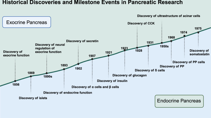
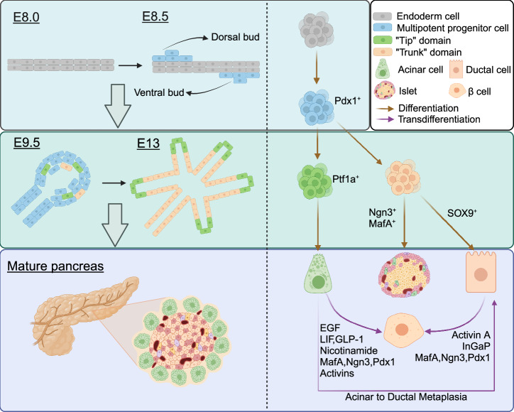
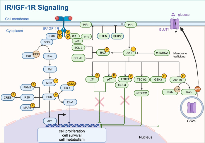
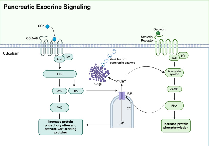
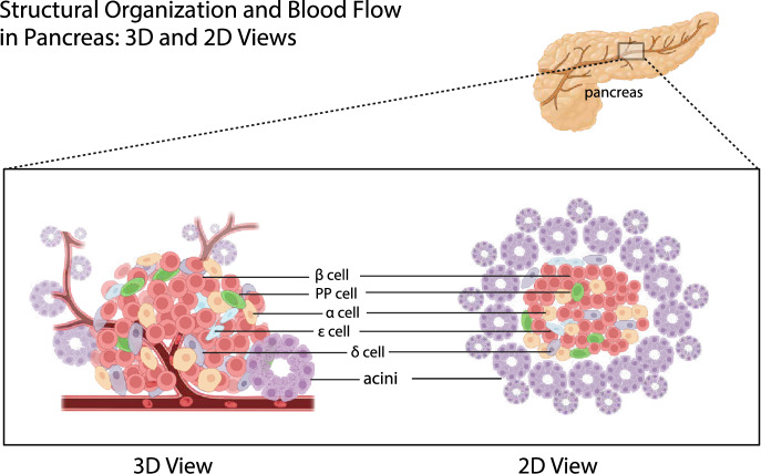
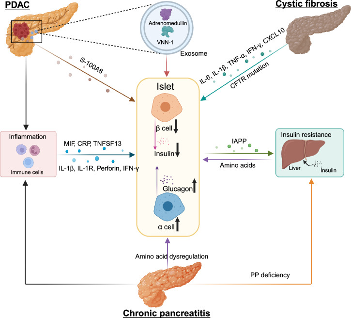
The pancreas, an organ with dual functions, regulates blood glucose levels through the endocrine system by secreting hormones such as insulin and glucagon. It also aids digestion through the exocrine system by secreting digestive enzymes. Complex interactions and signaling mechanisms between the endocrine and exocrine functions of the pancreas play a crucial role in maintaining metabolic homeostasis and overall health. Compelling evidence indicates direct and indirect crosstalk between the endocrine and exocrine parts, influencing the development of diseases affecting both. From a developmental perspective, the exocrine and endocrine parts share the same origin-the "tip-trunk" domain. In certain circumstances, pancreatic exocrine cells may transdifferentiate into endocrine-like cells, such as insulin-secreting cells. Additionally, several pancreatic diseases, including pancreatic cancer, pancreatitis, and diabetes, exhibit potential relevance to both endocrine and exocrine functions. Endocrine cells may communicate with exocrine cells directly through cytokines or indirectly by regulating the immune microenvironment. This crosstalk affects the onset and progression of these diseases. This review summarizes the history and milestones of findings related to the exocrine and endocrine pancreas, their embryonic development, phenotypic transformations, signaling roles in health and disease, the endocrine-exocrine crosstalk from the perspective of diseases, and potential therapeutic targets. Elucidating the regulatory mechanisms of pancreatic endocrine and exocrine signaling and provide novel insights for the understanding and treatment of diseases.


Conflict of interest statement

Competing interests: The authors declare no competing interests.

Figures

Fig. 1
Fig. 1
Historical discoveries and milestone events in pancreatic research. Significant historical milestones in the understanding of pancreatic structure and function highlight the discovery timeline of various components and functions of the pancreas. Created in BioRender.com
Fig. 2
Fig. 2
Pancreas development, cell differentiation, and transdifferentiation. At E8.5, the dorsal and ventral endoderm thickens to form two buds, called dorsal buds and ventral buds respectively. Cells in the two buds are Pdx1+ multipotent progenitor cells. At E9.5, the curled structure forms. Subsequently, these multipotent stem cells differentiate into two domains that together form a ramified structure. “Tip” domain cells differentiated into Ptf1a+ acinar precursor cells, while “Trunk” domain cells differentiated into SOX9+ duct precursor cells and Ngn3+ endocrine precursor cells. Transdifferentiation of acinar cells and ductal cells into islet endocrine cells and ADM can occur in mature pancreas under specific conditions. Created in BioRender.com
Fig. 3
Fig. 3
IR/IGF-1R signaling. The binding of insulin or IGF-1 to their receptors activates IRS proteins, leading to the activation of PI3K (p85/p110) and subsequent conversion of PIP2 to PIP3. Activation of AKT, which phosphorylates targets such as TSC1/2, mTORC1, AS160, and GSK3, regulating glucose uptake via GLUT4 translocation, protein synthesis, and cell survival. The Ras/MAPK pathway is activated through GRB2 and SOS, leading to the activation of RAF, MEK, and ERK, influencing cell proliferation and differentiation. The nuclear effects of these pathways include the regulation of transcription factors like FOXO, CREB, and Elk-1, affecting gene expression related to cell cycle, apoptosis, and metabolism. Created in BioRender.com
Fig. 4
Fig. 4
Pancreatic exocrine signaling. Two signaling pathways are involved in pancreatic exocrine function. Upon stimulation by acetylcholine or cholecystokinin (CCK), Gq protein-coupled receptors activate phospholipase C (PLC), which hydrolyzes phosphatidylinositol 4,5-bisphosphate (PIP2) into inositol triphosphate (IP3) and diacylglycerol (DAG). IP3 binds to its receptors (IP3R) on the endoplasmic reticulum (ER), triggering Ca2+ release into the cytoplasm. Increased intracellular Ca2+ concentration activates protein kinase C (PKC) and Ca2+-binding proteins, promoting enzyme secretion. Secretin binding to its receptor activates adenylate cyclase via Gs protein, increasing cAMP levels and activating protein kinase A (PKA), which also enhances enzyme secretion. This coordinated signaling ensures the efficient digestion of nutrients. Created in BioRender.com
Fig. 5
Fig. 5
Structural organization and blood flow in the pancreas: 3D and 2D views. The pancreas comprises various cell types, including acinar cells responsible for exocrine function and islet cells (α, β, δ, PP, and ε cells) responsible for endocrine function. Acinar cells are shown in clusters forming acini, while islet cells are scattered within the pancreatic tissue. The blood flow direction is indicated, starting from the branches of pancreaticoduodenal and splenic arteries entering the pancreas, first passing through the islets of Langerhans, and then flowing into the exocrine acinar and ductal cells. Created in BioRender.com
Fig. 6
Fig. 6
Mechanisms of PDAC-DM, CP-related DM, and CFRD. Inflammatory microenvironment is present in PDAC, CP, and CF. These diseases stimulate immune cells in the microenvironment to secrete various cytokines that act on β cells, causing β cell death or dysfunction. PDAC cells act on β cells through exosomes or their own secretions, causing them to be dysfunctional. β cells can secrete IAPP, mediating insulin resistance. CP can lead to amino acid disorders, resulting in abnormal numbers and dysfunction of β and α cells. CP can also mediate insulin resistance by reducing PP production. Some amino acids secreted by the liver act on α cells to secrete glucagon. The abnormal number and dysfunction of α and β cells is an important mechanism for the formation of diabetes mellitus. Created in BioRender.com

References

    1. Leung, P. S. Physiology of the pancreas. Adv. Exp. Med. Biol.690, 13–27 (2010). - PubMed
    1. Andersen, D. K. et al. Diabetes, pancreatogenic diabetes, and pancreatic cancer. Diabetes66, 1103–1110 (2017). - PMC - PubMed
    1. Pannala, R., Basu, A., Petersen, G. M. & Chari, S. T. New-onset diabetes: a potential clue to the early diagnosis of pancreatic cancer. Lancet Oncol.10, 88–95 (2009). - PMC - PubMed
    1. Malka, D. et al. Risk factors for diabetes mellitus in chronic pancreatitis. Gastroenterology119, 1324–1332 (2000). - PubMed
    1. Jolles, S. Paul Langerhans. J. Clin. Pathol.55, 243 (2002). - PMC - PubMed

MeSH terms

LinkOut - more resources