. 2025 Feb 13;15(1):5337.
doi: 10.1038/s41598-025-89930-0.

Intratumoral administration of mRNA COVID-19 vaccine delays melanoma growth in mice

Affiliations

Intratumoral administration of mRNA COVID-19 vaccine delays melanoma growth in mice

Dylan T Boehm et al. Sci Rep. .

Abstract

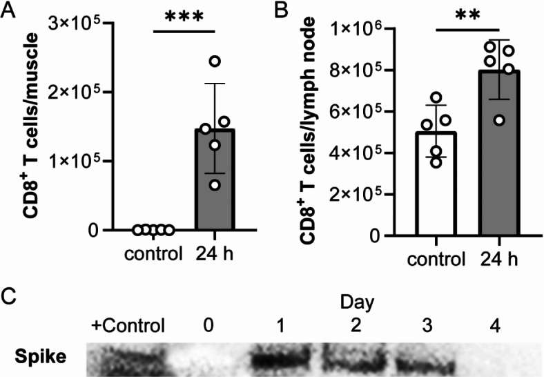
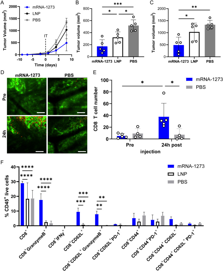
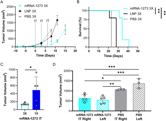
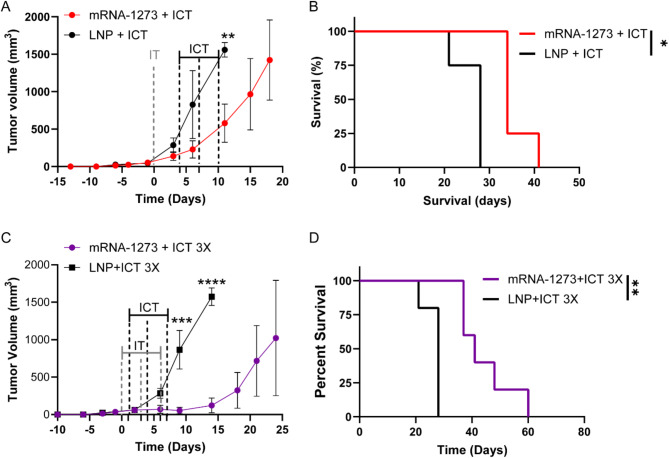
Immunotherapies are effective for cancer treatment but are limited in 'cold' tumor microenvironments due to a lack of infiltrating CD8+ T cells, key players in the anti-cancer immune response. The onset of the COVID-19 pandemic sparked the widespread use of mRNA-formulated vaccines and is well documented that vaccination induces a Th1-skewed immune response. Here, we evaluated the effects of an intratumoral injection of the mRNA COVID-19 vaccine in subcutaneous melanoma tumor mouse models. Tumor growth and survival studies following a single intratumoral injection of the COVID-19 vaccine showed significant tumor suppression and prolonged survival in established B16F10 subcutaneous tumor-bearing mice. mRNA vaccine treatment resulted in a significant increase in CD8+ T cell infiltration into the tumor microenvironment, as observed using intravital imaging and flow cytometry. Further tumor growth suppression was achieved using additional mRNA vaccine treatments. Combination administration of mRNA vaccine with immune checkpoint therapies demonstrated enhanced effects, further delaying tumor growth and improving the survival time of tumor-bearing mice. This study demonstrates that mRNA vaccines may be used as adjuvants for immunotherapies.

Keywords: COVID-19 vaccine; Immunotherapy; Melanoma; T cell infiltration; mRNA vaccine.


Conflict of interest statement

Declarations. Competing interests: The authors declare no competing interests.

Figures

Fig. 1
Fig. 1
mRNA-1273 vaccine immunization increases CD8 + T cell infiltration at the injection site and is uptaken by B16F10 melanoma cells. Flow cytometry of CD8+ T cells in the (A) muscle of injection site and (B) draining lymph node 24 h post immunization with 10 µg of mRNA-1273 or PBS control (n = 5 mice/group, n = 10 mice total). Data shown as mean (SD); unpaired student t test, **p = 0.0081, ***p < 0.001. (C) Cropped Western blot of spike protein in B16F10 cells at different timepoints after incubation with 200 µL (40 µg) mRNA-1273 (n = 3 independent experiments). Original full blots are presented in Supplementary Fig. 14.
Fig. 2
Fig. 2
mRNA-1273 vaccine intratumoral treatment reduces tumor growth and increases CD8+ T cells in the tumor. (A) B16F10 tumor growth curves and tumor volume measurements at (B) 4 days and (C) 8 days following a 20 µL intratumoral injection of 3 µg mRNA-1273, empty LNP or PBS (n = 5 mice/group, n = 15 mice total). Day 0 represents the time of IT injection when tumors reached 5 mm in diameter. (D) Representative intravital image of tumor (GFP, green) and CD8+ T cells (CD8a+, red) prior to (Pre) and 24 h post (24 h) 10 µL IT injection of 1.5 µg mRNA-1273 or PBS; scale bar represents 100 μm. (E) Corresponding quantification of the number of CD8+ T cells in the tumor (n = 5 mice treated with mRNA-1273, n = 4 mice treated with PBS, n = 9 mice total; n = 3 images per mouse per time point). Flow cytometry analysis of (F) CD8+ T cell subsets within the tumor 4 days post IT injection (n = 3 mice/group, n = 9 mice total). Data shown as mean (SD); one-way or two-way ANOVA followed by Tukey’s multiple comparison test, *p < 0.05, **p < 0.01, ***p < 0.001, ****p < 0.0001.
Fig. 3
Fig. 3
Enhanced tumor volume reduction results from using multiple mRNA-1273 IT injections. B16F10 (A) tumor growth curves and (B) survival curves following three 20 µL IT injections of 3 µg mRNA-1273, empty LNP or PBS (n = 5 mice treated with mRNA-1273 and LNP, n = 3 mice treated with PBS, n = 13 mice total); hazard ration (HR) of mRNA-1273/LNP is 0.043 with log-rank (Mantel-Cox) test, **p = 0.0035, HR of mRNA-1273/PBS is 0.024 with log-rank (Mantel-Cox) test, **p = 0.0082 and HR of LNP/PBS is 0.16 with log-rank (Mantel-Cox) test, p > 0.5. (C) B16F10 tumor volume measurements at 8 days post IT injection comparing three doses of mRNA-1273 versus a single mRNA-1273 IT dose (single dose mRNA-1273 IT data is same dataset as shown in Fig. 2C, n = 5 mice/group, n = 10 mice total). Abscopal effects of mRNA-1273 was evaluated using mice implanted with tumors on the right and left flanks. (D) B16F10 tumor volume measurements of treated right and untreated left flank tumors at day 8 post IT injection using three 20 µL IT injections of 3 µg mRNA-1273 or PBS (n = 5 mice treated with mRNA-1273 and n = 3 mice treated with PBS, n = 8 mice total). Data shown as mean (SD); unpaired student t test or two-way ANOVA followed by Tukey’s multiple comparison test, *p < 0.05, **p < 0.01, ***p < 0.001.
Fig. 4
Fig. 4
Enhanced therapeutic effects of mRNA-1273 and immune checkpoint therapy (ICT). B16F10 (A) tumor growth curves and (B) survival curves following a single 20 µL IT injections of 3 µg mRNA-1273 or empty LNP combined with ICT 4 days later (n = 4 mice/group, n = 8 mice total); *p = 0.043, **p = 0.0016. PBS + ICT control group reached tumor endpoint before ICT treatments were completed. HR of mRNA-1273 + ICT/LNP + ICT is 0.050 with log-rank (Mantel-Cox) test, *p = 0.011. B16F10 (C) tumor growth curves (*** p < 0.001, ****p < 0.0001) and (D) survival curves following 3 doses of a 20 µL IT injections of 3 µg mRNA-1273 or empty LNP followed by ICT 1 day later (n = 5 mice/group, n = 10 mice total); HR of mRNA-1273 + ICT 3X/LNP + ICT 3X is 0.043 with log-rank (Mantel-Cox) test, *p = 0.0039. Tumor growth data shown as mean (SD), unpaired student t test comparing tumor volume post treatment.
Fig. 5
Fig. 5
IT injection of mRNA-1273 does not alter cytokines or have transcriptional effects. (A) Cytokine levels of tumor, inguinal lymph node, and sera at 48 h after IT injection with 3 µg mRNA-1273 (mRNA) or PBS. Heat map shown depicts mean pg/ml of each cytokine log10 transformed (n = 5 mice/group, n = 10 mice total). tSNE plot of (B) immune cell types separated by treatment groups and (C) the cells defined by their single-cell transcriptome analysis from combined mRNA-1273 and PBS IT injections. (D) The proportions of cell projections in tumors treated with mRNA-1273 or PBS IT injections. (BD) scRNAseq analysis was performed using pooled samples (n = 5 mice/group, n = 10 mice total), prior to library preparation.

References

    1. Raskov, H., Orhan, A., Christensen, J. P. & Gögenur, I. Cytotoxic CD8 + T cells in cancer and cancer immunotherapy. Br. J. Cancer. 124, 359–367. 10.1038/s41416-020-01048-4 (2021). - PMC - PubMed
    1. Bos, R. & Sherman, L. A. CD4 + T-Cell help in the Tumor Milieu is required for recruitment and cytolytic function of CD8+ T lymphocytes. Cancer Res.70, 8368–8377. 10.1158/0008-5472.can-10-1322 (2010). - PMC - PubMed
    1. Thomas, N. E. et al. Tumor-infiltrating lymphocyte Grade in primary melanomas is independently associated with melanoma-specific survival in the population-based genes, environment and melanoma study. J. Clin. Oncol.31, 4252–4259. 10.1200/jco.2013.51.3002 (2013). - PMC - PubMed
    1. DONIZY, P. et al. Paucity of Tumor-infiltrating lymphocytes is an unfavorable prognosticator and predicts Lymph Node metastases in cutaneous melanoma patients. Anticancer Res.35, 351–358 (2015). - PubMed
    1. Zhao, Y. et al. A leukocyte infiltration score defined by a gene signature predicts melanoma patient prognosis. Mol. Cancer Res.17, 109–119. 10.1158/1541-7786.mcr-18-0173 (2019). - PMC - PubMed

Publication types

MeSH terms