. 2025 Mar;57(3):668-679.
doi: 10.1038/s41588-025-02081-w. Epub 2025 Feb 13.

Active repression of cell fate plasticity by PROX1 safeguards hepatocyte identity and prevents liver tumorigenesis

Affiliations

Active repression of cell fate plasticity by PROX1 safeguards hepatocyte identity and prevents liver tumorigenesis

Bryce Lim et al. Nat Genet. 2025 Mar.

Abstract

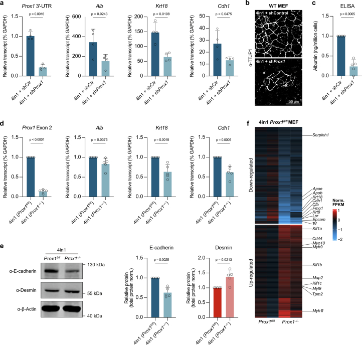
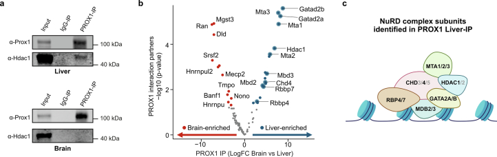
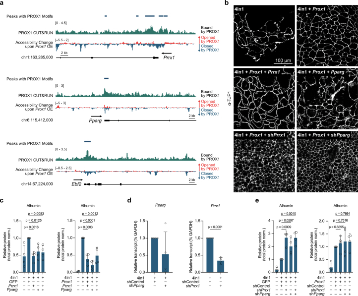
Cell fate plasticity enables development, yet unlocked plasticity is a cancer hallmark. While transcription master regulators induce lineage-specific genes to restrict plasticity, it remains unclear whether plasticity is actively suppressed by lineage-specific repressors. Here we computationally predict so-called safeguard repressors for 18 cell types that block phenotypic plasticity lifelong. We validated hepatocyte-specific candidates using reprogramming, revealing that prospero homeobox protein 1 (PROX1) enhanced hepatocyte identity by direct repression of alternative fate master regulators. In mice, Prox1 was required for efficient hepatocyte regeneration after injury and was sufficient to prevent liver tumorigenesis. In line with patient data, Prox1 depletion caused hepatocyte fate loss in vivo and enabled the transition of hepatocellular carcinoma to cholangiocarcinoma. Conversely, overexpression promoted cholangiocarcinoma to hepatocellular carcinoma transdifferentiation. Our findings provide evidence for PROX1 as a hepatocyte-specific safeguard and support a model where cell-type-specific repressors actively suppress plasticity throughout life to safeguard lineage identity and thus prevent disease.


Conflict of interest statement

Competing interests: The authors declare no competing interests.

Figures

Fig. 1
Fig. 1. Safeguard repressor screen.
a, Schematic representation of safeguard repressor prediction based on TF expression and DNA-binding motif analysis. b, Scores of top safeguard repressor candidates across 18 cell types, including lifelong expression, repressor/activator activity, tumor suppressor roles and TFs promoting indicated cell fate (asterisks). c, TF expression and motif presence analysis highlight top six hepatocyte safeguard repressor candidates. d, Prox1 expression (left) and motif counts in signature genes of indicated cell types (right). e, log-rank test between Kaplan–Meier curves from patients with HCC in TCGA, segregated by high versus low expression of indicated candidates. f, Bulk RNA-seq expression of hepatocyte candidates during mouse liver development. g, Validation of top three liver candidates by overexpression using 4-in-1 iHep reprogramming. h, TJP1 immunofluorescence of cells in g (n = 3). i, Quantification of TJP1+ cells and albumin secretion of cells in g (n = 3). Scale bar = 100 µm (h). Bar and line graphs show mean (n = 3), and error bars = s.d. (f,i). Two-tailed Dunnett’s test (i). P values are displayed. TCGA, The Cancer Genome Atlas; iHep, induced hepatocyte; RPKM, reads per kilobase million; WB, western blot; IF, immunofluorescence. Source data
Fig. 2
Fig. 2. PROX1 suppresses liver cancer formation and progression.
a, PROX1 expression in tumors from patients with HCC and paired normal tissue. b, PROX1 protein in patients with HCC liver tumors and adjacent nontumor tissues (n = 15). c, Survival of patients with HCC stratified by PROX1 expression levels (40% high-expression cutoff). d, Survival of patients with HCC ranked by PROX1 amplification status. e, Confluency of Hep3B cancer cells upon PROX1 shRNA-KD or OE for 7 days normalized to uninduced controls (n = 3). f, Differential ATAC–seq accessibility in Hep3B cells upon PROX1 OE for 2 days (n = 3; adjusted P < 0.05). g, Prox1 and hepatocyte signature expression across 7,793 single cells in healthy (day 0) and MYC-induced mouse HCC model (day 28). h, Mouse livers following HDTVI-mediated Myc OE and Trp53 KO with constitutive Prox1 OE (n = 5). i, Percentage of GFP+ tumors in mice treated as in h (n = 4). j, Survival of mice treated as in h following constitutive Prox1 OE (n = 5) or doxycycline-inducible late Prox1 OE (n = 4) at day 14 compared to control (n = 5 for constitutive OE and n = 3 for late OE). k, HDTVI-induced liver tumors following Kras(G12D) OE and Trp53 KO with constitutive Prox1 OE (OE; n = 4). l, Percentage of GFP+ tumors in mice treated as in k (n = 4). m, Survival of mice treated as in k following constitutive Prox1 OE or control (n = 4). Scale bar = 10 mm (h,k). Bar graphs and scatter plots show mean, error bars = s.d., boxplots show median and IQR and whiskers = 1.5× IQR from specified replicates. Unpaired two-tailed t test (a,b,i,l), log-rank test (c,d,j,m) and two-tailed one-sample t test (e). P values are displayed. ROI, region of interest; HR, hazard ratio; KO, knockout; KD, knockdown; OE, overexpression; IQR, interquartile range. Source data
Fig. 3
Fig. 3. PROX1 is necessary during liver regeneration.
a, Schematic representation of DDC diet-induced liver injury and regeneration in mice. The figure is created with BioRender.com. b, Pseudotime-ordered RNA expression of Prox1 and hepatocyte signature of mice treated as in a, across 1,866 single cells. c, Liver injury and recovery as in a following Cre-mediated Prox1 deletion in conditional Prox1fl/fl knockout mice. d, Number of HNF4+ cells per area in livers from mice treated as in c (n = 3). e, Serum levels of ALP from mice treated as in c (n = 3). Bar graphs show mean (n = 3), error bars = s.d., and unpaired two-tailed t test. P values are displayed. Source data
Fig. 4
Fig. 4. PROX1 promotes hepatocyte cell fate via multilineage repression.
a, Hepatocyte reprogramming time course with or without Prox1 OE analyzed by single-cell RNA-seq. b, A total of 22,761 cells treated as in a, following clustering and UMAP projection (n = 2). c, Annotation of cells in b based on experimental treatment and time point. d, Projection of hepatocyte and fibroblast identity onto cells in b. e, Hepatocyte and fibroblast identity scores in iHep cluster from b. f, Correlation of various cell identity scores with 4-in-1 or PROX1 regulon activity. g, Immunofluorescence of reprogrammed hepatocytes (4-in-1), neurons (Ascl1) or myocytes (Myod1) with or without Prox1 OE stained for TJP1 (hepatocyte), TUBB3 (neuronal) or desmin (myocyte) at day 14 (n = 3). h, Immunofluorescence quantification of cells in g (n = 3). i, Prox1 KO during iHep reprogramming via Cre-mediated deletion in Prox1fl/fl MEFs. j, TJP1 immunofluorescence of cells in i) at day 14 (n = 3). k, Number of TJP1+ cells (n = 3) and albumin secretion (n = 5) of cells in i. l, Proportion of reprogrammed cells in i, positive for desmin or TJP1 (n = 3). Scale bar = 100 µm (g,j). Bar graphs show mean, error bars = s.d., boxplots show median and IQR, whiskers = 1.5× IQR, from specified replicates. and unpaired two-tailed t test. P values are displayed. Source data
Fig. 5
Fig. 5. Direct repression of alternative fates by PROX1.
a, PROX1 interaction partners by mass spectrometry upon immunoprecipitation from mouse liver (n = 4). b, TJP1 immunofluorescence upon 4-in-1 iHep reprogramming with indicated PROX1 DBD fusion constructs at day 14 (n = 3). c, Number of TJP1+ cells (n = 3) and albumin secretion (n = 5) of cells in b. d, Proportion of reprogrammed cells in b, positive for indicated cell-type markers and morphology (n = 3). e, RNA-seq of differentially expressed genes from cells in b compared to DBD as a control at day 7 (n = 2). f, Percent overlap of upregulated and downregulated genes in e (n = 2). Scale bar = 100 µm (b). Bar graphs show mean from specified replicates, and error bars = s.d. Two-tailed Dunnett’s test (c), two-tailed Fisher’s exact test (f) and P values are displayed. FPKM, fragments per kilobase million; NS, not significant. Source data
Fig. 6
Fig. 6. Alternative master regulators are silenced by PROX1.
a, Hepatocyte reprogramming time course with or without Prox1 OE clustered based on RNA-seq expression (n = 2) and ATAC–seq accessibility (n = 3) displayed as scaled log(FC) compared to control. b, Overlap of cell signature markers with genes in clusters from a (adjusted P < 0.01). c, Enrichment or depletion of TF targets in promoters of genes clustered in a (adjusted P < 0.05). d, Prediction of key TFs downstream of PROX1 in a. e, Pearson correlation of indicated TF levels and expression of their target genes in a predict activator versus repressor activity. f, TJP1 immunofluorescence of 4-in-1 iHep reprogramming with OE or shRNA-mediated KD of Prrx1 or Pparg at day 14 (n = 3). g, Albumin western blot quantification of cells in f normalized to controls (n = 5). h, Proposed PROX1 gene regulatory network in iHep reprogramming. Scale bar = 100 µm (f). Bar graphs show mean, boxplots show median and IQR and whiskers = 1.5× IQR from specified replicates. Benjamini–Hochberg-adjusted two-tailed Fisher’s exact test (b,c), unpaired two-tailed t test (g) and P values are displayed. Source data
Fig. 7
Fig. 7. PROX1 regulates HCC versus CCA fate trajectories.
a, PROX1 expression in patients with HCC and CCA. The figure is created with BioRender.com. b, Pearson correlation of indicated markers with PROX1 expression in patients from a. c, Immunohistology of HCC (Myc/Trp53) and CCA (Akt/Notch) mice at the endpoint following Prox1 KD or OE compared to control (n = 5). d, Quantification of KRT19+ and HNF4+ cells in GFP+ tumors from c (n = 5, multiple regions per liver). e, Selected differentially expressed genes following RNA-seq from mice in (c) Prox1 KD (n = 2) or Late Prox1 OE (n = 2–3). Scale bar = 50 µm (c). Bar graphs and scatter plots show mean from specified replicates, error bars = s.d., unpaired two-tailed t test and P values are displayed. HE, hematoxylin and eosin. Source data
Extended Data Fig. 1
Extended Data Fig. 1. In silico and reprogramming screen identifies safeguard repressors.
a, Single-cell t-SNE of 18 cell types annotated by the Tabula Muris consortium. b, Cell-type-specific gene signatures used in this study displayed across all cells in a. c, Equations to calculate a safeguard repressor score (SRS) for each TF (see Methods for details). d, Safeguard repressor score analysis of 1,296 TFs shows neuronal candidates, including MYT1L. e, Myt1l expression and number of MYT1L motifs in cell-type-signature promoters. f, RNA-seq expression of the top neuronal safeguard repressor candidates in mouse brain development. g, Odds ratio of PROX1 CUT&RUN peaks from mouse liver in cell-type-signature gene promoters. h, TCGA survival curves for patients with HCC stratified by expression (high/low) of indicated safeguard repressors. i, Western blot of FLAG-tagged candidates upon overexpression at day 2 during hepatocyte reprogramming (n = 3). j, Western blot of albumin and E-cadherin at day 7 of hepatocyte reprogramming with indicated candidates (n = 3). k, Quantification of albumin and E-cadherin protein expression in j, normalized to total protein expression (n = 3). l, Expression analysis of indicated hepatocyte markers in cells treated as in j using qRT-PCR. Bar graphs show mean (n = 3), error bars = SD, two-tailed Dunnett’s test (k,l) or log-rank test (h). p values are displayed. Source data
Extended Data Fig. 2
Extended Data Fig. 2. PROX1 induces chromatin closure and growth delays in mouse HCC cell lines.
a, PROX1 protein level in liver sections from patients with HCC (n ≥ 3). b,c, Inducible PROX1 overexpression (OE; b) and knockdown (shPROX1; c) in Hep3B cells at day 2 compared to control (Ctr) by qRT-PCR (n = 6 each). d, MA-plot of ATAC-seq differentially accessible regions (DARs) upon PROX1 OE in Hep3B cells at day 2 (n = 3). e, PCA of DARs in d labeled by condition and replicate. f, Genomic annotations of chromatin peaks closed upon PROX1 OE compared to control determined by GREAT. g, IGV tracks of PROX1 binding by CUT&RUN and ATAC-seq accessibility change at the MYC locus in Hep3B cells 2 days after PROX1 OE compared to control (n = 3 each). h, GSEA normalized enrichment scores (NES) for MYC targets and apoptosis upon inducible PROX1 OE for 7 days in Hep3B cells or 2 days in Myc/Trp53 HCC mouse models (day 16 collection; n = 3 each). i, Doxycycline dose-dependent Prox1 OE in mouse tumor-derived cell lines transformed with Trp53 KO with OE of Myc (Myc/Trp53; n = 2) or Kras(G12D) (Kras/Trp53; n = 3) determined by western blot at day 3. j, Quantification of PROX1 protein levels of cells treated as in i. k, Confluency of cells treated as in i normalized to uninduced controls. Scale bar = 100 µm (a). Bar graphs show mean with error bars = SD, boxplots show median and interquartile range (IQR), whiskers = 1.5× IQR, from indicated biological replicates. Two-sided t test (b,c), unpaired t test (j) or one-sample t test (k). P values are displayed. Source data
Extended Data Fig. 3
Extended Data Fig. 3. PROX1 prevents liver cancer induction and progression in mice.
a, HCC-like tumor induction by Myc OE and Trp53 KO using HDTVI with constitutive Prox1 OE or GFP controls. b, Hematoxylin and eosin (H&E), HNF4 and KRT19 histology of livers treated as in a (n ≥ 3). c, Tumor numbers upon treatment as in a, following OE of Prox1-IRES-GFP (n = 3) vs GFP control (n = 4). d, Size of tumors (cross-sectional area) in c with or without GFP expression indicating transgene expression. e, Schematic of doxycycline-inducible late Prox1 OE at day 14 following HDTVI-tumor induction as in a. f, Livers treated as in e following histological staining for HNF4 and CASP3 at day 16 (n ≥ 3). g, Quantification of CASP3 levels in GFP+ tumors in f following late Prox1 OE (n = 3) vs GFP control (n = 4). h, GFP staining in livers treated as in e at the endpoint. i, Quantification of GFP+ tumor nodule numbers shown in h (n = 4). j, Western blot of PROX1 following PGK- and EF1a-promoter driven OE in vivo and in vitro compared to controls. k, Mouse livers following HDTVI to induce Myc OE and Trp53 KO with Prox1 OE using PGK- or EF1a-promoters (n = 4) compared to PGK-GFP controls (n = 5). l, Overall survival of mice treated as in k. m, Tumor induction via HDTVI-mediated Kras(G12D) OE and Trp53 KO with constitutive Prox1 OE or GFP controls. n, Liver histology following treatment as in m stained with H&E, HNF4 and KRT19 (n ≥ 3). o, Tumor numbers upon treatment as in m following OE of Prox1-IRES-GFP (n = 4) vs GFP control (n = 4). p, Size of tumors (cross-sectional area) in o with or without GFP expression indicating transgene expression. Scale bars = 10 mm, 5 mm and 500 µm (k,b,f,h,n). Bar graphs show mean values from indicated biological replicates, error bars = SD. Unpaired two-tailed t test (c,d,g,i,o,p), Bonferroni correction (d,p) and log-rank test (l). p values are displayed. The figure is created with BioRender.com. Source data
Extended Data Fig. 4
Extended Data Fig. 4. Impaired regeneration upon DDC-liver injury in Prox1-deleted mice.
a, Mouse liver histology stained for PROX1 and GFP 2 weeks following AAV GFP-Cre-mediated Prox1 deletion in conditional Prox1 knockout mice (Prox1fl/fl) compared GFP-ΔCre control mice before injury induction (n = 3). b, H&E, HNF4, SOX9 and KRT19 liver staining treated as in a and following 2 weeks of DDC diet and 2 weeks of recovery with normal diet (n = 3). c, Percentage of HNF4+ and KRT19+ cells in liver sections from mice treated as in b (n = 3). d, Serum levels of aspartate transaminase and alanine aminotransferase from mice treated as in b (n = 3). Scale bar = 5 mm and 500 µm (a,b). Bar graphs show mean (n = 3), error bars = SD and unpaired two-sided t test (c,d). P values are displayed. Source data
Extended Data Fig. 5
Extended Data Fig. 5. Depletion of Prox1 reduces hepatocyte reprogramming efficiency and fidelity.
a, Expression of Prox1 and hepatocyte markers at day 14 of iHep reprogramming upon Prox1 or control shRNA treatment based on qRT-PCR. b,c, TJP1 immunofluorescence (b) and normalized albumin secretion (c) of cells in a. d, Expression of indicated genes at day 14 of iHep reprogramming using Prox1fl/fl MEFs upon treatment with Cre (Prox1−/−) or ΔCre (Prox1fl/fl) based on qRT-PCR. e, E-cadherin (hepatocyte) and desmin (muscle) protein quantification of cells in d based on western blot analysis. f, Differential gene expression of cells in d based on RNA-seq (n = 2). Scale bar = 100 µm (b). Bar graphs show mean of n = 4 (a,b) or n = 5 (ce) biological replicates, error bars = SD and two-tailed t test. p values are displayed. Source data
Extended Data Fig. 6
Extended Data Fig. 6. PROX1 interacts with the repressive NuRD complex in liver but not in hippocampus.
a, Immunoprecipitation (IP) of PROX1 from primary mouse liver and hippocampus compared to IgG control followed by western blot using indicated antibodies (n = 4 each). b, Mass spectrometric identification and analysis of differential PROX1 interaction partners between hippocampus and liver (n = 4 each; Methods). c, Cartoon of the repressive NuRD complex highlighting liver-specific PROX1 interaction partners in black. The figure is created with BioRender.com. Source data
Extended Data Fig. 7
Extended Data Fig. 7. PROX1 predominantly closes bound chromatin and silences associated genes.
a, MA-plot of ATAC-seq differentially accessible regions (DAR) at day 2 of iHep reprogramming with or without Prox1 OE (n = 3; Methods). b, Percentage of closed and opened regions upon Prox1 OE in a. c, PCA of DARs in a labeled by condition and replicate and upon OE of GFP or Prox1 alone in MEFs (n = 3). d, Correlation of DARs between indicated conditions and replicates in c. e, Top: mean Tn5 transposon adapter insertions at indicated conditions from a centered at PROX1 CUT&RUN binding sites. Bottom: chromatin accessibility changes at PROX1 binding sites between 4-in-1 + GFP and 4-in-1 + Prox1 indicate decreased accessibility upon Prox1 OE. f, Differential gene expression of cells in a based on RNA-seq (n = 2), for genes with differentially accessible promoter (p adj < 0.05) within ±2 kb of their TSS and an overlapping CUT&RUN peak with a PROX1 binding motif (Methods). Source data
Extended Data Fig. 8
Extended Data Fig. 8. Prrx1 and Pparg are key PROX1 target genes during iHep reprogramming.
a, IGV tracks of PROX1 CUT&RUN chromatin binding (n = 3) and ATAC-seq accessibility (n = 3) change at day 2 of iHep reprogramming with or without Prox1 OE at indicated target promoters. b, TJP1 immunofluorescence at day 14 of iHep reprogramming with indicated combinations of TF OE and/or shRNA-mediated KD treatments (n = 4). c, Albumin western blot levels at day 14 of iHep reprogramming with OE of Prrx1 or Pparg and co-overexpression of GFP or Prox1 (n = 4). d, Expression of Pparg and Prrx1 upon shRNA-KD at day 14 of iHep reprogramming determined by qRT-PCR. e, Albumin western blot levels at day 14 of iHep reprogramming upon Prrx1 or Pparg KD with or without Prox1 OE (n = 4). Scale bar = 100 µm (b). Bar graphs show means normalized to total protein levels in c and e or GAPDH expression (d; n = 4), error bars = SD, two-tailed Dunnett’s test (c,e) or two-tailed t test (d). P values are displayed. Source data
Extended Data Fig. 9
Extended Data Fig. 9. PROX1 loss can enhance HCC liver tumor formation in mice.
a, Overall survival of patients with HCC and high PROX1 expression (40% expression cutoff) compared to CCA. b, Representative mouse livers at endpoint of HCC induction by Myc OE and Trp53 KO using HDTVI with PX330-sgRNA-Prox1 KO compared to control (n ≥ 3). c, Number of tumors upon treatment as in b following Prox1 KO vs control (Ctr; n = 3). d, Survival curve of HCC mice treated as in b comparing Prox1 KO (n = 4) with control (n = 3). e, Mouse livers at endpoint of HCC modeling as in b with EF1a-GFP-shProx1 KD compared to control (n ≥ 3). f, Number of tumor nodules with diameter >0.2 mm treated as in e at day 14 (n = 5). g, Tumor size (cross-sectional area) treated as in e in tumors with or without GFP expression (n = 5). h, Tumor numbers upon treatment as in e (n = 5). i, Survival curve of HCC mice treated as in e (n = 5). Scale bar = 10 mm (b,e). Bar graphs show mean from specified biological replicates, error bars = SD, log-rank test (a,d,i), unpaired two-tailed t test (c,g,h) and Bonferroni correction (g). P values are displayed. Source data
Extended Data Fig. 10
Extended Data Fig. 10. PROX1 can shift liver tumor fate from CCA to HCC in mice.
a, CCA liver tumor induction via HDTVI-mediated Akt and Notch1 receptor intracellular domain (NICD) (Akt/Notch) OE with constitutive Prox1 OE compared to controls. b, Mouse livers at endpoint of CCA induction as in a (n ≥ 3). c, Quantification of GFP+ tumors following constitutive OE of Prox1-IRES-GFP (n = 4) vs GFP as in a (n = 5). d, Tumors size (cross-sectional area) in c with or without GFP expression indicating transgene expression. e, Number of tumors upon treatment as in c. f, Survival curve of CCA mice treated as in c. log-rank test. g, Schematic of doxycycline-inducible late Prox1 OE at day 21 following HDTVI-CCA tumor induction as in a. h, Mouse livers 1–2 weeks following late Prox1 OE induced in CCA model as in g (n ≥ 3). i, Histology of livers treated as in g, stained with H&E, KRT19, HNF4 and SOX9 (n = 4). j, Quantification of KRT19 and SOX9 (CCA marker) and HNF4 (HCC marker) positive cells in GFP+ tumors following late Prox1 OE (n = 4) vs GFP (n = 4) from g. Scale bar = 10 mm and 50 µm (b,h,i). Bar graphs show mean from specified biological replicates, error bars = SD and unpaired two-sided t test. P values are displayed. The figure is created with BioRender.com. Source data

References

    1. Sánchez Alvarado, A. & Yamanaka, S. Rethinking differentiation: stem cells, regeneration, and plasticity. Cell157, 110–119 (2014). - PMC - PubMed
    1. Hanahan, D. Hallmarks of cancer: new dimensions. Cancer Discov.12, 31–46 (2022). - PubMed
    1. Balsalobre, A. & Drouin, J. Pioneer factors as master regulators of the epigenome and cell fate. Nat. Rev. Mol. Cell Biol.23, 449–464 (2022). - PubMed
    1. Garcia-Bellido, A. in Ciba Foundation Symposium 29—Cell Patterning (eds Porter, R. & Rivers, J.) Ch. 8 (Ciba Foundation, 1975).
    1. Lewis, E. B. A gene complex controlling segmentation in Drosophila. Nature276, 565–570 (1978). - PubMed

MeSH terms

Substances