Multicenter Study
. 2025 Feb 13;13(1):29.
doi: 10.1186/s40478-025-01946-9.

Blood biomarker fingerprints in a cohort of patients with CHRNE-related congenital myasthenic syndrome

Affiliations
Multicenter Study

Blood biomarker fingerprints in a cohort of patients with CHRNE-related congenital myasthenic syndrome

Adela Della Marina et al. Acta Neuropathol Commun. .

Abstract

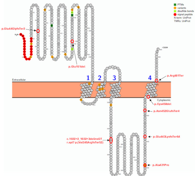
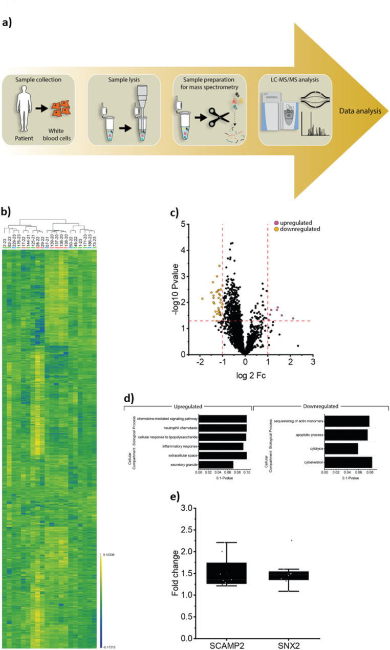
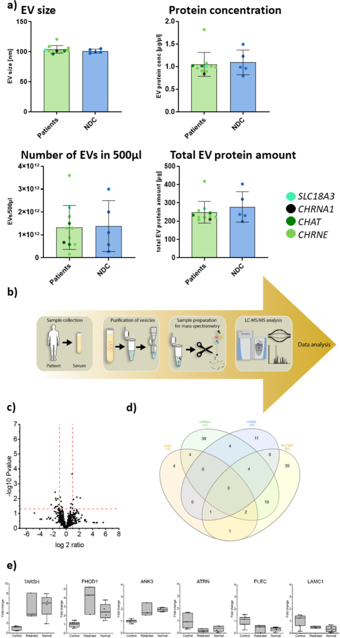
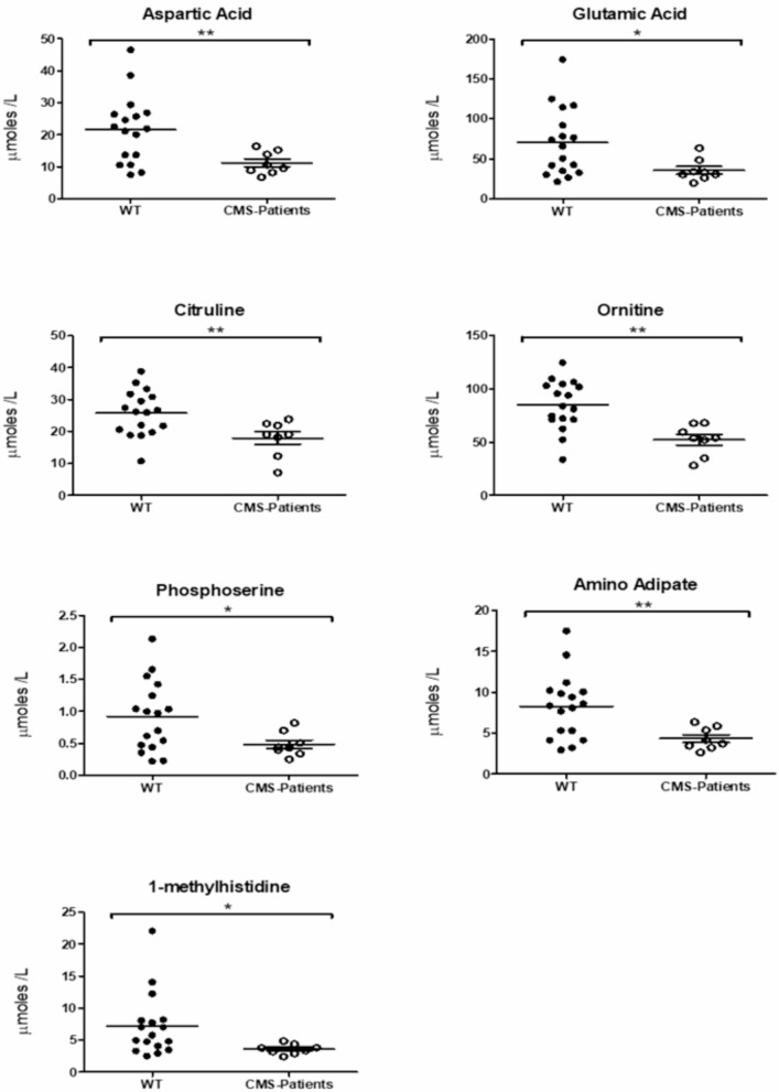
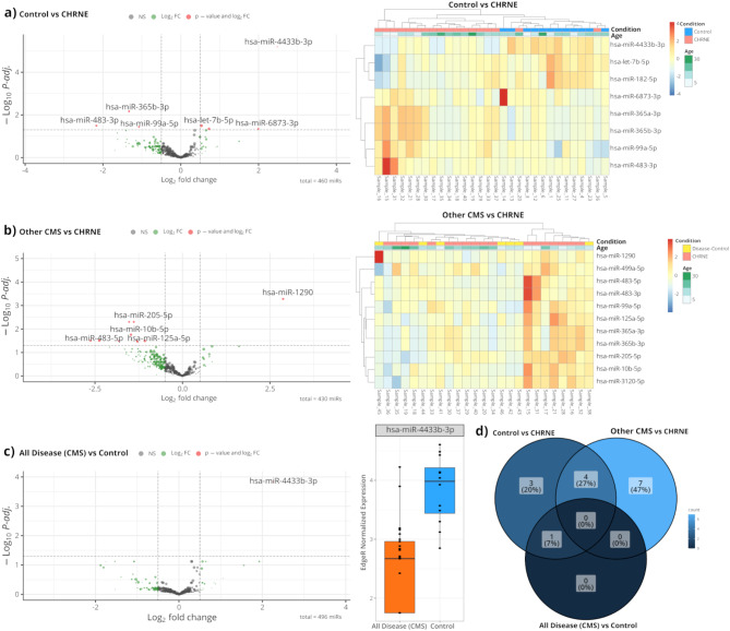
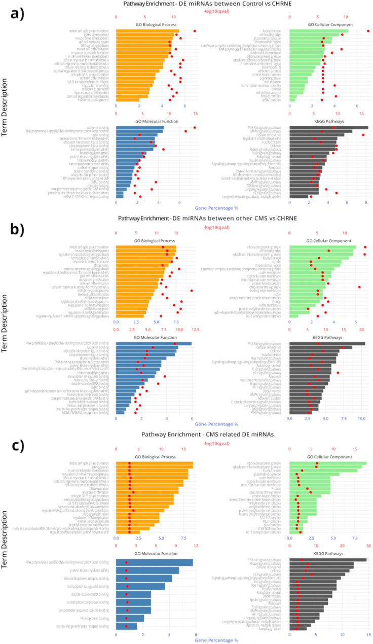
Mutations in CHRNE encoding the epsilon subunit of acetylcholine receptor result in impaired neuromuscular transmission and congenital myasthenic syndrome (CMS) with variying severity of symptoms. Although the pathophysiology is well-known, blood biomarker signatures enabling a patient-stratification are lacking. This retrospective two-center-study includes 19 recessive CHRNE-patients (AChR deficiency; mean age 14.8 years) from 13 families which were clinically characterized according to disease severity. 15 patients were classified as mildly and 4 patients as moderate to severely affected. Seven known pathogenic and one unreported variant (c.1032 + 2_1032 + 3delinsGT) were identified. Biomarker discovery was carried out on blood samples: proteomics was performed on white blood cells (WBC; n = 12) and on extracellular vesicles (EV) purified from serum samples (n = 7) in addition to amino acid profiling (n = 9) and miRNA screening (n = 18). For miRNA studies, 7 patients with other CMS-subtypes were moreover included. WBC-proteomics unveiled a significant increase of 7 and a decrease of 36 proteins. In silico studies of these proteins indicated affection of secretory granules and the extracellular space. Comparison across patients unveiled increase of two vesicular transport proteins (SCAMP2 and SNX2) in severely affected patients and indeed EV-proteomics revealed increase of 7 and decrease of 13 proteins. Three of these proteins (TARSH, ATRN & PLEC) are known to be important for synaptogenesis and synaptic function. Metabolomics showed decrease of seven amino acids/ amino acid metabolites (aspartic and glutamic acids, phosphoserine, amino adipate, citrulline, ornithine, and 1-methyhistidine). miRNA-profiling showed increase miR - 483 - 3p, miR-365a-3p, miR - 365b - 3p and miR-99a, and decrease of miR-4433b-3p, miR-6873-3p, miR-182-5p and let-7b-5p in CHRNE-patients whereas a comparison with other CMS subtypes showed increase of miR - 205 - 5p, miR - 10b - 5p, miR-125a-5p, miR-499-5p, miR-3120-5p and miR - 483 - 5p and decrease of miR - 1290. Our combined data introduce a molecular fingerprint on protein, metabolic and miRNA level with some of those playing different roles along the neuromuscular axis.

Keywords: CMS biomarkers; CMS extracellular vesicles; CMS metabolites; CMS miRNA; CMS white blood cells.


Conflict of interest statement

Declarations. Ethics approval and consent to participate: Our study was approved by the ethical committee of the University Hospital Duisburg-Essen (19-9011-BO) and for Spanish patients by ethical comitee of the Institut de Recerca Sant Joan de Deu (PIC-147-23). The study was conducted in accordance with the principles of the Declaration of Helsinki. Consent for publication: All participians or their representatives have given consent for publication of their clinical data as part of the above named studies. Competing interests: The authors declare no competing interests.

Figures

Fig. 1
Fig. 1
Schematic overview of distribution of CHRNE-variants identified in our patients across the protein structure. Protter: interactive protein feature visualization and integration with experimental proteomic data [46]
Fig. 2
Fig. 2
Study of protein signature of white blood cells derived from CHRNE-patients. (a) Schematic representation of proteomic workflow applied on white blood cells derived from CHRNE-patients and healthy controls. (b) Proteingenic changes are shown in the heatmap on the individual protein level. (c) Volcano plot depicting statistically significant dysregulated proteins in white blood cells of CHRNE-patients. Increased proteins are represented by purple dots wereas decreased proteins are represented by organge dots. (d) GO-term based in silico analysis of pathways affected by protein dysregulations affected by upregaulated and downregulated proteins, respectively. (e) Box plots highlighting the increase of SCAMP2 and SNX2 as two proteins relevant for vesicular transport in cells derived from CHRNE-patients compared to controls
Fig. 3
Fig. 3
Study of EVs purified from blood derived of CHRNE-related CMS patients. (a) Characterization of EVs purified from sera derived from CMS-patients and age-matched controls including determination of size, protein concentration number per 500 µl and total protein amount. (b) Schematic representation of proteomic workflow applied on EVs derived from CMS patients and healthy controls. (c) Volcano plot depicting statistically significant dysregulated proteins in EVs of CHRNE-patients. Increased proteins are represented by purple dots wereas decreased proteins are represented by organge dots. (d) Venn diagram showing overlaps of dysregulated EV proteins across the different CMS subtypes. (e) Box plots depicting TARSH, ATRN and PLEC as three proteins of major interest in addition to ANK3, LAMC1 and FHOD1 dysregulated in EVs of CHRNE-patients. No changes of these proteins were noted between patients with and without intellectual disability
Fig. 4
Fig. 4
Study of metabolic status in sera derived from CHRNE-patients. Mass spectrometry unveils statistically significant decrease of aspartic and glutamic acids, phosphoserine, amino adipate, citrulline, ornithine & 1-methyhistidine
Fig. 5
Fig. 5
Differentially expressed miRNAs (DEmiRNAs) between CHRNE-patients, other CMS subtype patients and healthy controls. Volcanos and heatmaps of the top DEmiRNAs based on the adjusted p value (< 0.05) for (a) CHRNE-patients versus controls and (b) CHRNE-patients versus other CMS subtypes. In heatmaps, the CHRNE group is shown in red colour, the control group (healthy individuals) is shown in blue colour and the other CMS subtype group in yellow. The colour key panel shows the Z-score values calculated for each miRNA, by subtracting the row-mean and then dividing by the standard deviation. Z-scores describe the expression of each miRNA in relation to the mean. Overexpressed miRNAs are shown in red, underexpressed miRNAs in blue. White colour indicates expression change close to 0. Hierarchical clustering was performed for samples and miRNAs. (c) Comparison of CHRNE-patients and other CMS subtypes versus controls revealed only miR-4433b-3p to be differentially expressed. (d) Venn diagram showing the number of unique and common miRNAs that reached significace in the three comparisons, CHRNE-patients versus controls, CHRNE-patients versus other CMS subtypes, CHRNE-patients and other CMS subtypes versus controls
Fig. 6
Fig. 6
Enriched biological processes based on the experimentally validated target genes of the DEmiRNAs. Gene target of the DEmiRNAs and pathway analysis of the experimentally validated target genes of the differentially expressed miRNAs between (a) CHRNE-patients versus controls, (b) CHRNE- patents versus other CMS subtype patients. (c) Functional analysis of the experimentally validated target genes of the DEmiRNAs in both comparisons, CHRNE-patients versus controls and CHRNE-patients versus other CMS subtype patients. For the functional analysis, the top 20 enriched ontologies from GO biological processes, GO cellular components, GO molecular function and KEGG pathways are present

Similar articles

References

    1. Ohkawara B, Ito M, Ohno K (2021) Secreted signaling molecules at the neuromuscular junction in physiology and pathology. Int J Mol Sci Int J Mol Sci; p. 1–16 - PMC - PubMed
    1. Engel AG, Shen XM, Selcen D, Sine SM (2015) Congenital myasthenic syndromes: Pathogenesis, diagnosis, and treatment. Lancet Neurol. Lancet Publishing Group, pp 420–434 - PMC - PubMed
    1. Abicht A, Muller Jj LochmüllerH (2017) Congenital Myasthenic Syndromes - GeneReviews®- NCBI Bookshelf
    1. Abicht A, Stucka R, Karcagi V, Herczegfalvi A, Horváth R, Mortier W et al (1999) A common mutation (ε1267delG) in congenital myasthenic patients of Gypsy ethnic origin. Neurology 53:1564–1569 - PubMed
    1. Richard P, Gaudon K, Haddad H, Ammar AB, Genin E, Bauché S et al (2008) The CHRNE 1293insG founder mutation is a frequent cause of congenital myasthenia in North Africa. Neurology 71:1967–1972 - PubMed

Publication types

LinkOut - more resources