Review
. 2025 Jan 14;17(1):e77418.
doi: 10.7759/cureus.77418. eCollection 2025 Jan.

Pulmonary Manifestations in Patients With Hematologic Malignancies: In Pursuit of an Accurate Diagnosis

Affiliations
Review

Pulmonary Manifestations in Patients With Hematologic Malignancies: In Pursuit of an Accurate Diagnosis

Jose C Alvarez-Payares et al. Cureus. .

Abstract

Pulmonary involvement is common in patients with hematologic malignancies (HMs) and varies depending on the underlying condition, including lymphoproliferative disorders, acute leukemia, myelodysplastic syndrome, and allogeneic stem cell transplantation. Pulmonary complications are a frequent cause of morbidity and mortality in these patients, often resulting from the immunosuppressive effects of the disease or its treatment. The clinical manifestations of these complications are nonspecific, and their differential diagnosis is broad, encompassing both infectious and noninfectious causes. A thorough clinical assessment requires consideration of factors such as the patient's history, baseline immune status, treatment regimens, time since the last chemotherapy, and environmental exposures. Radiographic imaging, particularly high-resolution CT, plays a critical role in evaluating these complications, helping clinicians identify distinct patterns of pulmonary involvement. Therefore, a personalized diagnostic approach is essential, and multidisciplinary management is crucial for optimal patient care.

Keywords: complications; diagnostic imaging; differential diagnosis; hematologic neoplasms; mortality; pulmonary disease.


Conflict of interest statement

Conflicts of interest: In compliance with the ICMJE uniform disclosure form, all authors declare the following: Payment/services info: All authors have declared that no financial support was received from any organization for the submitted work. Financial relationships: All authors have declared that they have no financial relationships at present or within the previous three years with any organizations that might have an interest in the submitted work. Other relationships: The authors of this article reside in two different countries, because Dr. Edwin Uriel Suárez completed his studies in Internal Medicine at the University of Antioquia, Medellín, but subsequently moved to Spain to pursue his studies in Hematology. Nevertheless, the authors have maintained contact for the preparation of this article.

Figures

Figure 1
Figure 1. Primary mediastinal NHL
A 24-year-old female patient with primary mediastinal large B-cell lymphoma. A heterogeneous mass is observed in the anterior mediastinum (blue arrow), extending to the medial mediastinum. Bilateral pleural effusion with right predominance is noted (red arrow), along with lingular atelectasis (yellow arrow). NHL, non-Hodgkin lymphoma Image courtesy: Pulmonology Service of the Internal Medicine Department, University of Antioquia. Consent for open access publication was obtained from the patient.
Figure 2
Figure 2. Adenopathies and pulmonary nodules
An 18-year-old patient with HL, nodular sclerosis variant. A lymph node conglomerate is observed toward the aortopulmonary window (blue arrow), with various nodular opacities in the lung parenchyma due to invasion by proximity (red arrow). HL, Hodgkin lymphoma Image courtesy: Pulmonology Service of the Internal Medicine Department, University of Antioquia. Consent for open access publication was obtained from the patient.
Figure 3
Figure 3. Consolidations
A 30-year-old male patient with classic HL and relapses of high-grade B-cell lymphoma, NOS. Neoplastic involvement of the mediastinum is observed (blue arrow), along with bilateral pleural effusion (red arrow) and right lung consolidations (yellow arrows). HL, Hodgkin lymphoma; NOS, not otherwise specified Image courtesy: Pulmonology Service of the Internal Medicine Department, University of Antioquia. Consent for open access publication was obtained from the patient.
Figure 4
Figure 4. Lymphangitic carcinomatosis
A 43-year-old patient with a medical history of common variable immunodeficiency and HL. Mild pleural and pericardial effusion is observed (blue arrow), along with widespread interlobular septal thickening suggesting lymphangitic carcinomatosis (red arrow). HL, Hodgkin lymphoma Image courtesy: Pulmonology Service of the Internal Medicine Department, University of Antioquia. Consent for open access publication was obtained from the patient.
Figure 5
Figure 5. Leukemia lung involvement
A 27-year-old male patient with the common phenotype of ALL. Chest CT reveals peripheral ground-glass opacities in the upper left lobe, along with central acinar micronodules of random distribution (blue arrow). Infectious causes were ruled out through BAL. ALL, acute lymphoblastic leukemia; BAL, bronchoalveolar lavage Image courtesy: Pulmonology Service of the Internal Medicine Department, University of Antioquia. Consent for open access publication was obtained from the patient.
Figure 6
Figure 6. Pulmonary involvement in CLL
A 55-year-old male patient with a medical history of CLL. Bilateral peribronchovascular ground-glass opacities are observed (blue arrows). Pulmonary cytology from BAL was positive for lymphoid infiltration. BAL, bronchoalveolar lavage; CLL, chronic lymphocytic leukemia Image courtesy: Pulmonology Service of the Internal Medicine Department, University of Antioquia. Consent for open access publication was obtained from the patient.
Figure 7
Figure 7. EMH
A 28-year-old female patient with IgG lambda MM. Paravertebral and left-sided soft tissue thickening (blue arrow), a finding associated with EMH. EMH, extramedullary hematopoiesis; MM, multiple myeloma Image courtesy: Pulmonology Service of the Internal Medicine Department, University of Antioquia. Consent for open access publication was obtained from the patient.
Figure 8
Figure 8. Pulmonary leukostasis
A 39-year-old female patient with acute B-cell lymphoblastic leukemia and hyperleukocytosis. Chest CT reveals thickening of the bronchial walls (blue arrow), thickening of the interlobular septa (red arrow), and subpleural ground-glass opacities (yellow arrow). Image courtesy: Pulmonology Service of the Internal Medicine Department, University of Antioquia. Consent for open access publication was obtained from the patient.
Figure 9
Figure 9. Pulmonary embolism
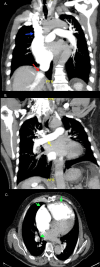
A 60-year-old male patient with severe pulmonary hypertension and JAK2-positive PV. The following findings were observed in chest CT angiography: (A) Dilatation of the superior vena cava (blue arrow) with contrast reflux into the inferior vena cava and opacification of the hepatic veins, indicative of right-sided heart failure (red arrow). (B) Dilation of the pulmonary artery (yellow arrow). (C) Prominence of right-sided cavities (green arrow), with eccentric filling defects involving lobar and segmental arteries, suggestive of chronic pulmonary embolism (green arrowhead). PV, polycythemia vera Image courtesy: Pulmonology Service of the Internal Medicine Department, University of Antioquia. Consent for open access publication was obtained from the patient.
Figure 10
Figure 10. Pulmonary edema
A 42-year-old female patient with a blastic crisis due to CML. Chest imaging reveals moderate bilateral pleural effusion associated with bilateral posterior-basal passive atelectasis (blue arrows), smooth thickening of the interlobular septa, and symmetrical bilateral ground-glass opacities suggestive of fluid overload (red arrow). CML, chronic myeloid leukemia Image courtesy: Pulmonology Service of the Internal Medicine Department, University of Antioquia. Consent for open access publication was obtained from the patient.
Figure 11
Figure 11. Cryptogenic organizing pneumonia
A 53-year-old female patient with splenic marginal zone lymphoma. Chest CT reveals subpleural and peribronchial consolidations, along with multifocal ground-glass opacities in the posterior segment of the upper right lobe and lateral segment of the middle lobe (blue arrow). Image courtesy: Pulmonology Service of the Internal Medicine Department, University of Antioquia. Consent for open access publication was obtained from the patient.
Figure 12
Figure 12. Chylothorax
A 26-year-old female patient with primary mediastinal large B-cell lymphoma. Chest imaging reveals a mass in the anterior mediastinum (blue arrow) and moderate bilateral pleural effusion (red arrows). Pleural fluid studies confirm the presence of chylothorax. Image courtesy: Pulmonology Service of the Internal Medicine Department, University of Antioquia. Consent for open access publication was obtained from the patient.
Figure 13
Figure 13. Differentiation syndrome and opportunistic mycosis
A 22-year-old male patient with acute promyelocytic leukemia developed dyspnea after 10 days of ATRA therapy. Chest CT revealed central ground-glass opacities in the lung parenchyma, predominantly in the upper and middle lobes (blue arrows), raising suspicion for differentiation syndrome. In addition, pulmonary nodules and peribronchovascular consolidations were observed (red arrows), and galactomannan antigen was positive in the BAL. Treatment for suspected aspergillosis with voriconazole was initiated, resulting in clinical improvement. ATRA, all-trans retinoic acid; BAL, bronchoalveolar lavage Image courtesy: Pulmonology Service of the Internal Medicine Department, University of Antioquia. Consent for open access publication was obtained from the patient.
Figure 14
Figure 14. Bacterial pneumonia
A 59-year-old male with lambda MM. Chest CT reveals extensive bilateral pleural effusion (blue arrows), along with alveolar consolidation in the inferior right lobe due to pneumonic involvement (red arrow). Haemophilus influenzae was isolated in microbiological studies. MM, multiple myeloma Image courtesy: Pulmonology Service of the Internal Medicine Department, University of Antioquia. Consent for open access publication was obtained from the patient.
Figure 15
Figure 15. Fungal infection
A 28-year-old female patient with nongerminal center diffuse large B-cell lymphoma and a prior history of cryptococcal meningitis. Chest CT reveals multiple centrilobular nodules in the lung parenchyma (blue arrow), ground-glass opacities in both lungs (red arrow), and basal consolidation in the left lung (yellow arrow). Cryptococcus spp. was isolated from the BAL. BAL, bronchoalveolar lavage Image courtesy: Pulmonology Service of the Internal Medicine Department, University of Antioquia. Consent for open access publication was obtained from the patient.

Similar articles

Cited by

References

    1. Pulmonary disease in non-pulmonary malignancy. Cheng GS, Possick JD. Clin Chest Med. 2017;38:0. - PubMed
    1. Cancer incidence, mortality, years of life lost, years lived with disability, and disability-adjusted life years for 29 cancer groups from 2010 to 2019: a systematic analysis for the Global Burden of Disease Study 2019. Kocarnik JM, Compton K, Dean FE, et al. JAMA Oncol. 2022;8:420–444. - PMC - PubMed
    1. Acute pulmonary complications in patients with hematologic malignancies. Choi MH, Jung JI, Chung WD, et al. Radiographics. 2014;34:1755–1768. - PubMed
    1. Gut microbiota predict pulmonary infiltrates after allogeneic hematopoietic cell transplantation. Harris B, Morjaria SM, Littmann ER, et al. Am J Respir Crit Care Med. 2016;194:450–463. - PMC - PubMed
    1. Pulmonary manifestations of lymphoma: a pictorial essay. Angirish B, Sanghavi P, Jankharia B. Lung India. 2020;37:263–267. - PMC - PubMed

LinkOut - more resources