. 2025 Feb 12;13(2):e70182.
doi: 10.1002/ccr3.70182. eCollection 2025 Feb.

Cold Ceramic for Repairing Root Perforations: A Case Report

Affiliations

Cold Ceramic for Repairing Root Perforations: A Case Report

Ali Chamani et al. Clin Case Rep. .

Abstract

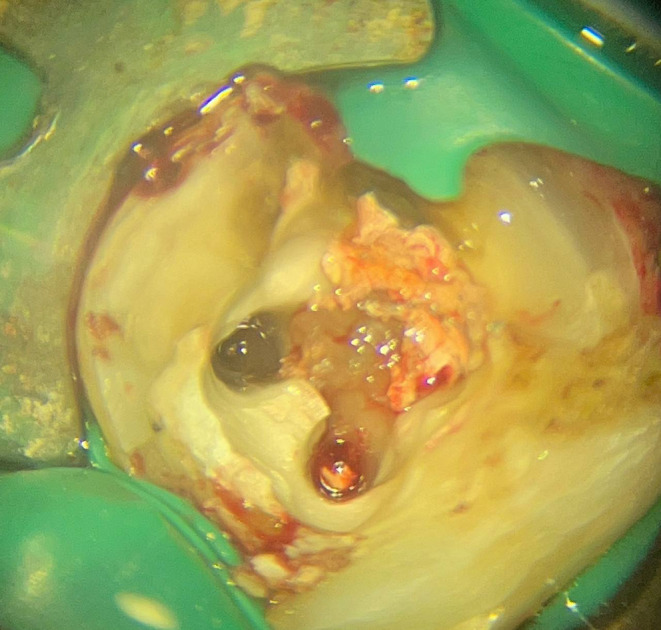
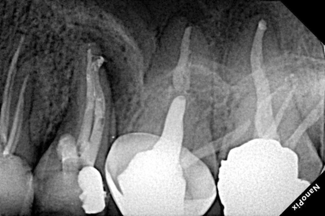
Root perforation may happen pathologically or iatrogenically at any stage of endodontic treatment. Repairing a Perforation can be challenging and can negatively affect the prognosis of the treatment. The prognosis depends on various factors, including the size and site of the perforation, the presence of microorganisms, and the time lapse between injury and repair. One crucial factor is using a suitable material with favorable biocompatibility, moisture tolerance, and sealing abilities. Thus, choosing the suitable perforation sealing material can significantly affect the outcomes. Many materials have been suggested for perforation repair. However, searching for an ideal material continues, indicating the complicated nature of root perforations. In this study, three patients, two male and one female, received treatment for root perforation, each varying in the prognosis determinant factors. All cases were managed non-surgically with cold ceramic (CC) as the repairing material. After follow-up, they were clinically and radiographically examined, and all three cases revealed relatively complete healing of the tissues and no signs or symptoms of inflammation. The results obtained from the presented cases indicated CC's sealing ability, biocompatibility, moisture tolerance, and bone and periodontium regeneration, which are essential for successful perforation repair. The favorable healing of the perforation and the elimination of inflammation in every case, as well as the existing literature, support the use of CC as a suitable material for sealing perforations. However, additional clinical research is recommended to further understand CC's qualities and potential.

Keywords: MTA; case report; cold ceramic; retreatment; root canal therapy; root filling material.


Conflict of interest statement

The authors declare no conflicts of interest.

Figures

FIGURE 1‐1
FIGURE 1‐1
Clinical examinations revealing abscess and deep pocket.
FIGURE 1‐2
FIGURE 1‐2
A periapical radiographic image showing the lesions around the mesial root.
FIGURE 1‐3
FIGURE 1‐3
After removing the filling material, massive bleeding was visible.
FIGURE 1‐4
FIGURE 1‐4
Clinical view of the perforations.
FIGURE 1‐5
FIGURE 1‐5
A periapical radiographic image showing the strip perforation.
FIGURE 1‐6
FIGURE 1‐6
Working length assessment and the apical perforation.
FIGURE 1‐7
FIGURE 1‐7
Repair of the strip perforation with CC.
FIGURE 1‐8
FIGURE 1‐8
A periapical radiographic image showing the #25 K‐file as the space maintainer.
FIGURE 1‐9
FIGURE 1‐9
A periapical radiographic image showing the 10‐mm deep pocket of tooth #30.
FIGURE 1‐10
FIGURE 1‐10
A periapical radiographic image showing the negotiation of the perforated canal to the estimated working length.
FIGURE 1‐11
FIGURE 1‐11
Repair of the perforations with CC.
FIGURE 1‐12
FIGURE 1‐12
Covering CC with Glass‐ionomer.
FIGURE 1‐13
FIGURE 1‐13
Postoperative radiograph.
FIGURE 1‐14
FIGURE 1‐14
Twelve‐month follow‐up examinations exhibiting tissue healing with no signs of inflammation.
FIGURE 1‐15
FIGURE 1‐15
Twelve‐month postoperative radiograph showing favorable healing of the lesions.
FIGURE 1‐16
FIGURE 1‐16
Clinical view of tooth #30 in the 15‐month follow‐up.
FIGURE 1‐17
FIGURE 1‐17
Fifteen‐month follow‐up examinations of tooth #30 with full cusp coverage PFM crown as the permanent restoration, healthy periodontium, negative BOP, and normal probing depth.
FIGURE 1‐18
FIGURE 1‐18
Periapical follow‐up radiographic image after 15 months of treatment, exhibiting complete tissue healing bone formation.
FIGURE 2‐1
FIGURE 2‐1
A periapical radiographic image showing the misposition of the file due to strip perforation.
FIGURE 2‐2
FIGURE 2‐2
Clinical examination of tooth #19 exhibiting swelling and abscess.
FIGURE 2‐3
FIGURE 2‐3
A periapical radiographic image demonstrateing furcal and coronal lesions.
FIGURE 2‐4
FIGURE 2‐4
Clinical view after removing the temporary filling material.
FIGURE 2‐5
FIGURE 2‐5
Clinical view after locating the actual orifices.
FIGURE 2‐6
FIGURE 2‐6
Negotiation and working length assessment.
FIGURE 2‐7
FIGURE 2‐7
Repair of the perforations with CC.
FIGURE 2‐8
FIGURE 2‐8
Postoperative radiograph.
FIGURE 2‐9
FIGURE 2‐9
Postoperative angled radiograph showing the furcal and strip perforations.
FIGURE 2‐10
FIGURE 2‐10
Clinical examinations of 3‐month follow‐up showing tissue healing with no signs of inflammation.
FIGURE 2‐11
FIGURE 2‐11
Three‐month follow‐up exhibiting favorable healing of the lesions.
FIGURE 2‐12
FIGURE 2‐12
Clinical examinations in the 11‐month follow‐up showing tooth #19 with full cusp coverage PFM crown as the permanent restoration and healthy periodontium.
FIGURE 2‐13
FIGURE 2‐13
Clinical examinations in the 11‐month follow‐up showing tooth #19 fully functional in the occlusion.
FIGURE 2‐14
FIGURE 2‐14
11‐month follow‐up exhibiting significant healing and tissue regeneration with no signs of inflammation.
FIGURE 3‐1
FIGURE 3‐1
Clinical examination revealing a deep pocket in the mesial of tooth #13.
FIGURE 3‐2
FIGURE 3‐2
A periapical radiograph image showing coronal and apical lesions due to mesial perforation and missed canal.
FIGURE 3‐3
FIGURE 3‐3
Clinical view of the mesial perforation.
FIGURE 3‐4
FIGURE 3‐4
Working length assessment.
FIGURE 3‐5
FIGURE 3‐5
Repair of the mesial perforation with CC.
FIGURE 3‐6
FIGURE 3‐6
Postoperative radiograph.
FIGURE 3‐7
FIGURE 3‐7
Clinical examination showing probing depth within normal limit, no bleeding on probing, and no signs of inflammation.
FIGURE 3‐8
FIGURE 3‐8
Eight‐month follow‐up shows nearly complete healing and tissue regeneration with no signs of inflammation.

References

    1. Clauder T., “Repair of Pulp Chamber and Root Perforations,” in Endodontic Advances and Evidence‐Based Clinical Guidelines (Wiley, 2022), 475–510.
    1. Estrela C., Decurcio D. A., Rossi‐Fedele G., Silva J. A., Guedes O. A., and Borges Á. H., “Root Perforations: A Review of Diagnosis, Prognosis and Materials,” Brazilian Oral Research 32, no. 1 (2018): e73, 10.1590/1807-3107bor-2018.vol32.0073. - DOI - PubMed
    1. Thamilselvan A. and Ramesh S., “Clinical Practice Guidelines for the Management of Endodontic Perforation,” Journal of Pharmacological Research 12 (2020): 4046–4053, 10.31838/ijpr/2020.SP2.501. - DOI
    1. Modaresi J., Parashos P., Mousavi R., Mirzaeeian A., and Almodaresi Z., “Treatment of Strip Perforation Using Cold Ceramic,” Dental Research Journal 20 (2023): 31. - PMC - PubMed
    1. Kakani A. K., Veeramachaneni C., Majeti C., Tummala M., and Khiyani L., “A Review on Perforation Repair Materials,” Journal of Clinical and Diagnostic Research 9, no. 9 (2015): ZE09, 10.7860/jcdr/2015/13854.6501. - DOI - PMC - PubMed

LinkOut - more resources