. 2025;2(1):8.
doi: 10.1038/s44328-024-00023-w. Epub 2025 Feb 11.

A same day α-synuclein RT-QuIC seed amplification assay for synucleinopathy biospecimens

Affiliations

A same day α-synuclein RT-QuIC seed amplification assay for synucleinopathy biospecimens

Sabiha Parveen et al. NPJ Biosens. 2025.

Abstract

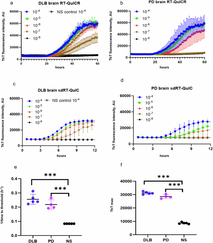
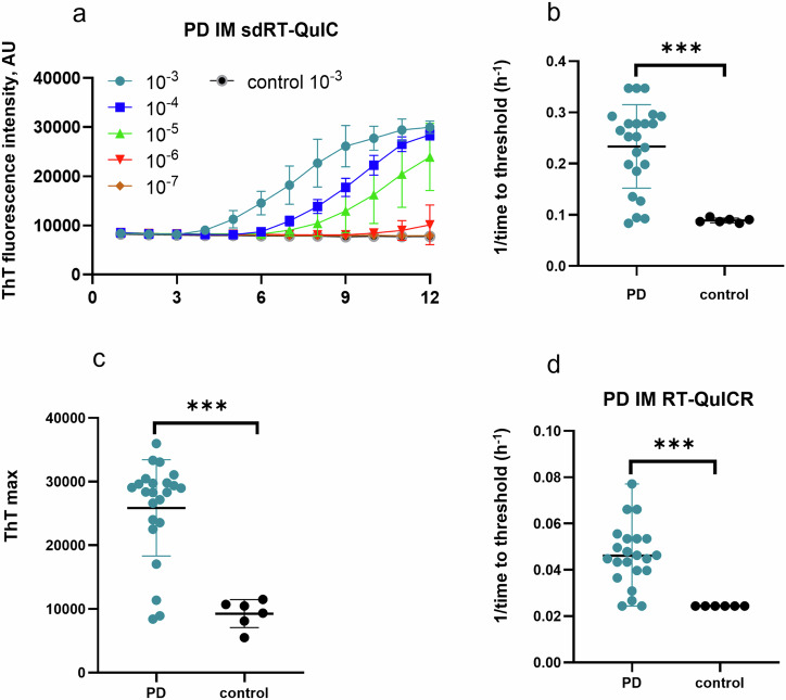
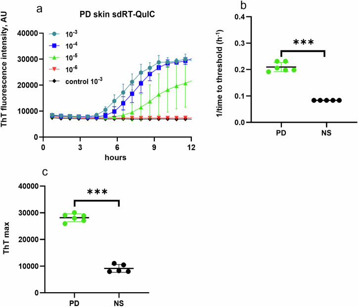
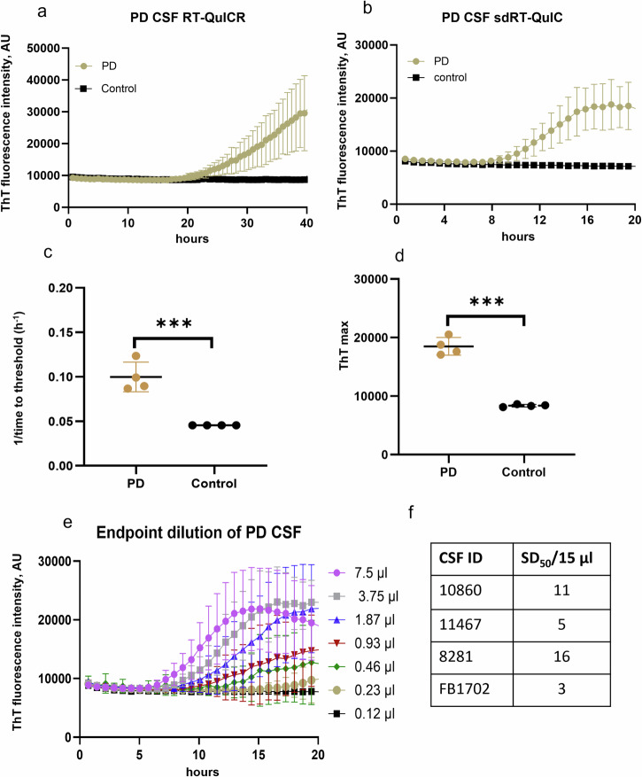
Parkinson's disease (PD), dementia with Lewy bodies (DLB), and other synucleinopathies are characterized by the accumulation of abnormal, self-propagating aggregates of α-synuclein. RT-QuIC or seed amplification assays are currently showing unprecedented diagnostic sensitivities and specificities for synucleinopathies even in prodromal phases years in advance of the onset of Parkinsonian signs or dementia. However, commonly used α-synuclein seed amplification assays take ≥48 h to perform as applied to patients' diagnostic biospecimens. Here, we report the development of a faster α-synuclein RT-QuIC assay that is as analytically sensitive as prior assays of this type, but can be completed in ≤12 h for brain, skin, and intestinal mucosa, with positive signals often arising in <5 h. CSF assays took a few hours longer. Our same-day α-synuclein RT-QuIC (sdRT-QuIC) assay should increase the practicality, cost-effectiveness, and throughput of measurements of pathological forms of α-synuclein for fundamental research, clinical diagnosis, and therapeutics development.

Keywords: Biochemistry; Biological techniques; Biomarkers.


Conflict of interest statement

Competing interestsB.C., C.O., and A.G.H. are inventors of patent applications pertaining to α-syn RT-QuIC technology. The other authors have declared that no competing interests exist.

Figures

Fig. 1
Fig. 1. Analysis of the brain using the RT-QuICR (a,b) and sdRT-QuIC (c-f) assays.
ad End-point dilution analyses of single DLB (a, c) and PD (b, d) specimens. ThT fluorescence traces show means ± SD of quadruplicate reactions at the designated dilutions given with respect to the original tissue mass rather than homogenates thereof. Non-synucleinopathy (NS) brain at 10−4 dilution was included as a negative control, with the traces shown in (a, c) also applying to the plots in (b, d). e 1/TTT and f ThT maxima were obtained using 10−4 DLB (n = 5), PD (n = 4), and NS (n = 5) brain dilutions. Data points represent means of quadruplicate reactions from individual cases, and bars represent the mean ± SD of the means from all cases of that type. The same brain specimens were used in these RT-QuICR and sdRT-QuIC assays. Statistical significance is denoted as *p < 0.05, **p < 0.01, ***p < 0.001.
Fig. 2
Fig. 2. sdRT-QuIC and RT-QuICR analyses of intestinal mucosa (IM) biopsies.
a Representative end-point dilution analysis of a PD and an NS control IM sample. Data points indicate means ± SD of quadruplicate reactions at the designated dilutions of PD (n = 1) and control IMs (n = 1). b 1/time to threshold and c ThT maxima obtained from 10-3 dilutions of PD (n = 23) and NS (n = 6) IMs. d 1/TTT at 10−3 dilution of the same panel of PD and NS IMs using the RT-QuICR assay (data summarized from Fig. 3 of ref. ). In bd, data points represent means of quadruplicate reactions from individual cases, and bars represent the mean ± SD of the means from all cases. The same IM specimens were used in the RT-QuICR and sdRT-QuIC assays. Statistical significance is denoted as *p < 0.05, **p < 0.01, ***p < 0.001.
Fig. 3
Fig. 3. sdRT-QuIC analysis of skin.
a End-point dilution analysis of skin samples from representative PD (n = 1) and NS (n = 1) cases. Data points indicate means ± SD of quadruplicate reactions seeded with the designated dilutions of the PD skin samples. b 1/TTT and c ThT maxima obtained from 10−3 dilutions of PD skin (n = 6) and NS control (n = 5) cases. In b, c, data points represent means of quadruplicate reactions from individual cases, and bars represent the mean ± SD of the means from all cases. Statistical significance is denoted as *p < 0.05, **p < 0.01, ***p < 0.001.
Fig. 4
Fig. 4. sdRT-QuIC analysis of CSF.
Representative analyses of single PD and NS control CSF specimens using a RT-QuICR and b sdRT-QuIC. Traces show means ± SD of ThT fluorescence from quadruplicate reactions. c 1/TTT and d ThT maxima within 20 h of sdRT-QuIC reactions seeded with PD (n = 4) and NS (n = 4) CSFs. The same CSF samples were used in the respective RT-QuICR and sdRT-QuIC assays. Data points represent means of quadruplicate reactions from individual cases, and bars represent the mean ± SD of the means from all cases. e End-point dilutions of a PD CSF by sdRT-QuIC. Each trace represents the mean ± SD ThT fluorescence of quadruplicate wells. f log SD50/15 µl values for PD CSFs (n = 4). Statistical significance is denoted as *p < 0.05, **p < 0.01, ***p < 0.001.
Fig. 5
Fig. 5. Seed concentration (log SD50/mg tissue) determination for DLB (n = 5) and PD (n = 4) brain, PD skin (n = 6), and PD IM (n = 23) samples by sdRT-QuIC.
Data points represent log SD50/mg tissue values calculated from sample dilution series with quadruplicate reactions at each dilution, and bars represent mean ± SD of values from all cases.

References

    1. Soto, C. Unfolding the role of protein misfolding in neurodegenerative diseases. Nat. Rev. Neurosci.4, 49–60 (2003). - PubMed
    1. Kraus, A., Groveman, B. R. & Caughey, B. Prions and the potential transmissibility of protein misfolding diseases. Annu. Rev. Microbiol.67, 543–564 (2013). - PMC - PubMed
    1. Caughey, B. & Lansbury, P. T. Jr Protofibrils, pores, fibrils, and neurodegeneration: separating the responsible protein aggregates from the innocent bystanders. Annu. Rev. Neurosci.26, 267–298 (2003). - PubMed
    1. Alam, P., Bousset, L., Melki, R. & Otzen, D. E. α‐synuclein oligomers and fibrils: a spectrum of species, a spectrum of toxicities. J. Neurochem.150, 522–534 (2019). - PubMed
    1. Koga, S., Sekiya, H., Kondru, N., Ross, O. A. & Dickson, D. W. Neuropathology and molecular diagnosis of synucleinopathies. Mol. Neurodegener.16, 83 (2021). - PMC - PubMed

LinkOut - more resources