. 2025 Mar;301(3):108306.
doi: 10.1016/j.jbc.2025.108306. Epub 2025 Feb 12.

The sterol-regulating human ARV1 binds cholesterol and phospholipids through its conserved ARV1 homology domain

Affiliations

The sterol-regulating human ARV1 binds cholesterol and phospholipids through its conserved ARV1 homology domain

Jessie Lee Cunningham et al. J Biol Chem. 2025 Mar.

Abstract

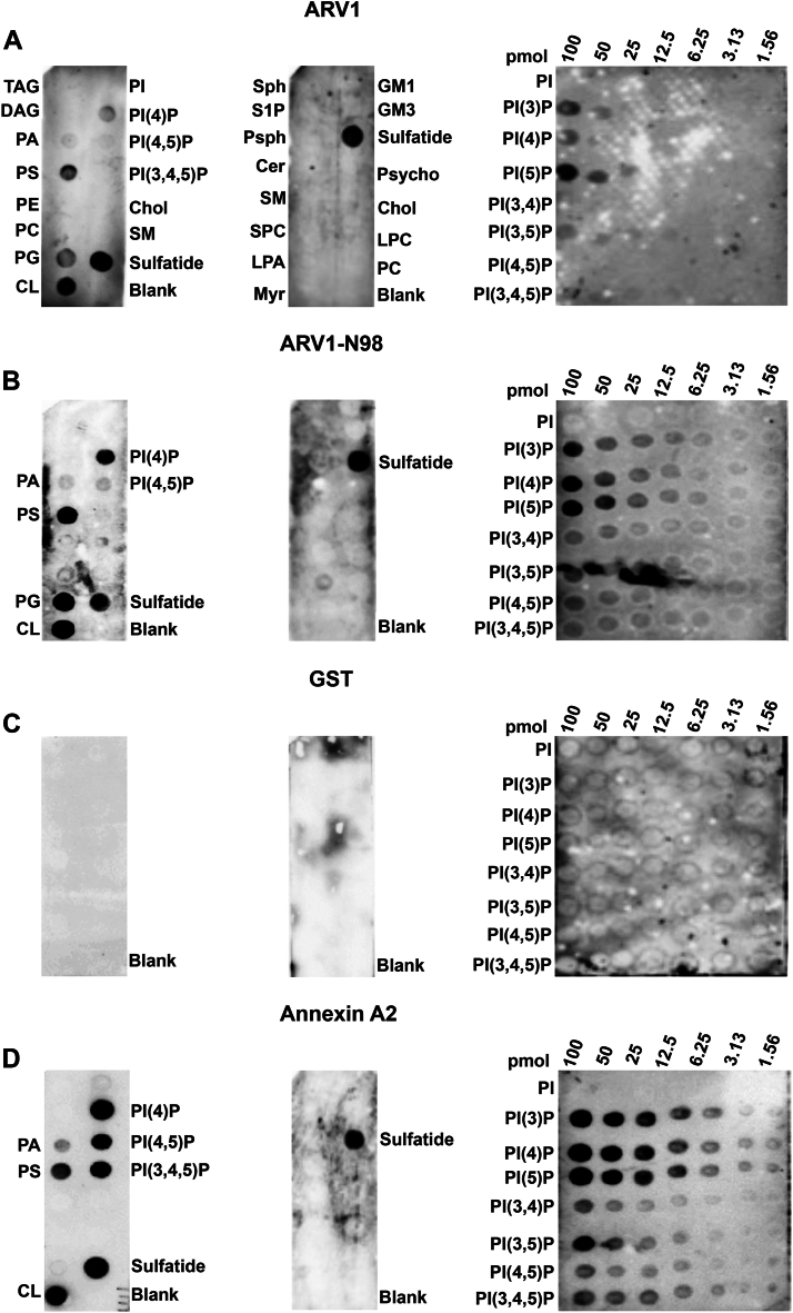
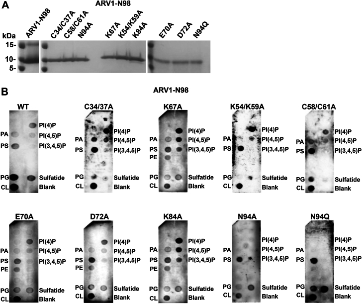
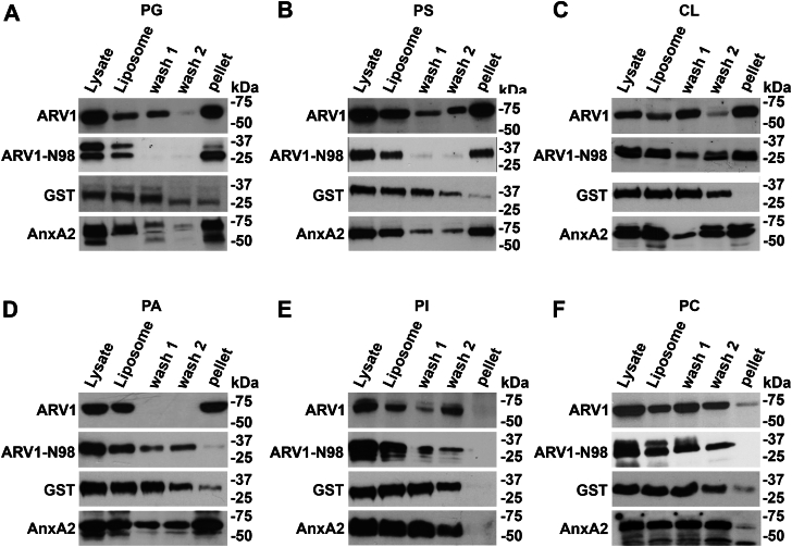
Evidence suggests that ARV1 regulates sterol movement within the cell. Saccharomyces cerevisiae cells lacking ScArv1 have defects in sterol trafficking, distribution, and biosynthesis. HepG2 cells treated with hARV1 antisense oligonucleotides accumulate cholesterol in the endoplasmic reticulum. Mice lacking Arv1 have a lean phenotype when fed a high fat diet and show no signs of liver triglyceride or cholesterol accumulation, suggesting a role for Arv1 in lipid transport. Here, we explored the direct lipid-binding activity of recombinant human ARV1 using in vitro lipid-binding assays. ARV1 lipid-binding activity was observed within the first N-terminal 98 amino acids containing the conserved ARV1 homology domain (AHD). The zinc-binding domain and conserved cysteine clusters within the AHD were necessary for lipid binding. Both full-length ARV1 and the AHD bound cholesterol, several phospholipids, and phosphoinositides with high affinity. The AHD showed the highest binding affinity for monophosphorylated phosphoinositides. Several conserved amino acids within the AHD were necessary for phospholipid binding. Biochemical studies suggested that ARV1 exists as a dimer in cells, with oligomerization being critical for ARV1 function, as amino acid mutations predicted to have a negative effect on dimerization caused weakened or complete loss of lipid binding. Our results show for the first time that human ARV1 can directly bind cholesterol and phospholipids. How this activity may function to regulate lipid binding and maintain proper lipid trafficking and/or transport in cells requires further studies.

Keywords: cholesterol-binding protein; lipid; lipid transport; lipid-binding protein; phosphoinositide; phospholipids.


Conflict of interest statement

Conflict of interest All authors are present or were past employees of Genesis Global Group, Inc.

Figures

Figure 1
Figure 1
Full-length ARV1 and N98 associate with cholesterol-containing liposomes. Phosphatidylcholine:cholesterol liposomes were generated as described in the “Experimental procedures” section. An aliquot of purified GST-tagged protein (lysate) was mixed with liposomes (liposome) and spun at 50,000g for 20 min. An aliquot of the supernatant was taken (wash 1) and the pellet was washed a second time. An aliquot of the second supernatant was taken (wash 2) and the pellet was resuspended (pellet). All washes and resuspensions of pellets were performed using floatation buffer. Proteins were resolved by SDS-PAGE and detected by Western blotting. A, Western blot of GST-ARV1 and GST-N98. B, Western blots of GST-ARV1 and GST-ARV1-N98 tagged protein binding to different concentration PC:Chol liposomes. Blots are representative images. (n = 5).
Figure 2
Figure 2
Full-length ARV1 and ARV1-N98 interact with phospholipids and phosphoinositides. Lipid overlay far westerns were performed using GST-ARV1, GST-ARV1-N98, GST, and GST-Annexin A2. A, GST-ARV1 lipid overlay interactions. B, GST-ARV1-N98 lipid overlay interactions. C, GST lipid overlay interactions. D, GST-Annexin A2 lipid overlay interactions. Blots are representative images. (n = 5).
Figure 3
Figure 3
The zinc-binding motif within the AHD is required for lipid–protein interaction.A, carboxy-terminal GST-ARV1 truncations were generated and used in lipid overlay far westerns. B, GST-ARV1-N70 lipid-binding interaction. C, GST-ARV1-N50 lipid-binding interaction. D, GST-ARV1-N30 lipid-binding interaction. Blots are representative images. (n = 5).
Figure 4
Figure 4
Amino acids comprising the human, rat, and mouse AHD. The zinc-binding domain is underlined. Identical amino acids are below. Conserved amino acids in bold were mutated.
Figure 5
Figure 5
Several conserved amino acids within the zinc-binding domain are required for specific lipid–protein interactions. Far westerns were performed using specific 6XHIS-ARV1-N98 mutant proteins (A). All amino acids residues were changed to alanine unless otherwise indicated. B, various site-directed mutants far westerns. Blots are representative images. (n = 5).
Figure 6
Figure 6
Full-length ARV1 and N98 proteins associate with phospholipid liposomes. Liposomes were made as described in figure legend 1. GST-ARV1, GST-ARV1-N98, GST, and GST-Annexin A2 were used for all experiments. A, protein binding to PG liposomes. B, protein binding to PS liposomes. C, protein binding to CL liposomes. D, protein binding to PA liposomes. E, protein binding to PI liposomes. F, protein binding to PC liposomes. GST was used as a control for nonspecific liposome association. Blots are representative images. (n = 5).
Figure 7
Figure 7
The Cys58/Cys61 cluster within the zinc-binding domain is required for N98 association with specific phospholipid-containing liposomes.A, GST-ARV1-N98 mutants were tested for liposome association. B, PG liposome binding. C, PA liposome binding. D, sulfatide-binding association. E, PI(4)P liposome binding. F, PE liposome binding. Blots are representative images. (n = 5).
Figure 8
Figure 8
N98 binds phospholipids with different affinities. HTRF assays were performed using 6XHIS-ARV1-N98 proteins. A constant concentration of 6XHIS-ARV1-N98 (0.75 μM) was used in the presence of increasing concentrations of biotinylated phospholipids. EC50 values were obtained using GraphPad Prism statistical analysis. A, 6XHIS-ARV1-N98 binding to biotinylated phospholipids. B, 6XHIS-N98 binding to biotinylated phosphoinositides.
Figure 9
Figure 9
Various phospholipids affect ARV1-lipid binding. For competition assays, 6XHIS-ARV1-N98 protein was held at a single concentration (0.75 μM) and a single biotinylated phospholipid was held at a constant concentration (80% maximum signal). Increasing concentrations of nonbiotinylated competitor lipids were added and IC50 values were obtained using GraphPad Prism statistical analysis. A, biotinylated-PG in the presence of the indicated nonbiotinylated PLs. B, biotinylated-PS in the presence of the indicated nonbiotinylated PLs. C, biotinylated-PA in the presence of the indicated nonbiotinylated PLs. D, biotinylated-CL in the presence of the indicated nonbiotinylated PLs. Statistical analysis is described in “Experimental procedures.”
Figure 10
Figure 10
ARV1 may form oligomers.A, nitrocellulose blots embedded with the indicated proteins were incubated with 6XHIS-ARV1 protein and subsequently probed with GST- or 6XHIS-antibodies. B, serial dilutions of yeast strains harboring various combinations of yeast two-hybrid ARV1/ARV1-N98 expression plasmids were grown on protein–protein interaction medium for 7 days. C, cells expressing various ARV1/ARV1-N98 NanoBRET plasmids were assayed for protein–protein interaction as described in “Experimental procedures.” Values are mean ± S.D. ∗∗∗p ≤ 0.0001. Blot is a representative image. (n = 5). Two way ANOVA was used for statistical analysis.
Figure 11
Figure 11
ARV1 may form a dimer in cells.A, Escherichiacoli expressed purified full-length ARV1 was run on a 2.5%-25% sucrose density gradient. Gradient fractions were resolved by SDS-PAGE and western analysis was performed using anti-ARV1 antibodies (inset). Molecular weight markers from left to right are ovalbumin (50 kDa), bovine serum albumin (77 kDa), and phosphorylase β (103 kDa). B, Western blot of E. coli lysates expressing ARV1-MYC or ARV1-FLG probed with anti-ARV1 antibodies. C, mixed lysates were immunoprecipitated with anti-FLAG antibodies; co-immunoprecipitated proteins were resolved by SDS-PAGE and transferred to nitrocellulose and probed with anti-MYC antibodies. D, mixed lysates were immunoprecipitated with anti-MYC antibodies; co-immunoprecipitated proteins were resolved by SDS-PAGE and transferred to nitrocellulose and probed with anti-FLAG antibodies. IgG antibodies were used as a control for nonspecific binding. Blots are representative images. (n = 5).
Figure 12
Figure 12
Certain conserved amino acids are required for lipid binding. The NanoBRET assay was performed with cells expressing either Halo-ARV1-Nluc-ARV1 (red bar), Halo-ARV1-Nluc-ARV1 (C58/C61A) (blue bar), Halo-ARV1-Nluc-ARV1 (K67A) (brown bar), or Halo-ARV1-Nluc-ARV1 (N94A) (purple bar) proteins. Halo-Nluc–expressing cells served as a negative control. Values are mean ± S.D. ∗∗∗p ≤ 0.0001. (n = 5). Two way ANOVA was used for statistical analysis.
Figure 13
Figure 13
Schematic diagram indicating the effects of various amino acid mutations on lipid binding. Schematic of the AHD of human ARV1. The zinc-binding domain is underlined. Conserved amino acids mutated are color coded with unique colors. In the case of double mutations, the color coding is the same. The arrows are the same color as the amino acids and represent whether lipid binding was increased (up arrow) or decreased (down arrow).

Similar articles

References

    1. Wong L.H., Copic A., Levine T.P. Advances on the transfer of lipids by lipid transfer proteins. Trends Biochem. Sci. 2017;42:516–530. - PMC - PubMed
    1. Ikonen E., Zhou X. Cholesterol transport between cellular membranes: a balancing act between interconnected lipid fluxes. Dev. Cell. 2021;56:1430–1436. - PubMed
    1. Quinville B.M., Deschenes N.M., Ryckman A.E., Walia J.S. A comprehensive review: sphingolipid metabolism and implications of disruption in sphingolipid homeostasis. Int. J. Mol. Sci. 2021;22:5793. - PMC - PubMed
    1. Dudek J. Role of cardiolipin in mitochondrial signaling pathways. Front. Cell Dev. Biol. 2017;5:90. - PMC - PubMed
    1. Lorent J.H., Levental K.R., Ganesan L., Rivera-Longsworth G., Sezgin E., Doktorova M., et al. Plasma membranes are asymmetric in lipid unsaturation, packing and protein shape. Nat. Chem. Biol. 2020;16:644–652. - PMC - PubMed

LinkOut - more resources