. 2025 Feb 14;23(1):181.
doi: 10.1186/s12967-024-05906-0.

Micheliolide attenuates sepsis-induced acute lung injury by suppressing mitochondrial oxidative stress and PFKFB3-driven glycolysis

Affiliations

Micheliolide attenuates sepsis-induced acute lung injury by suppressing mitochondrial oxidative stress and PFKFB3-driven glycolysis

Wenhan Li et al. J Transl Med. .

Abstract

Background: Sepsis is a potentially fatal condition with a significant risk of death. Acute lung injury (ALI) is a life-threatening complication of sepsis, and the inflammatory response plays a critical role in sepsis-induced ALI. The protective effects of micheliolide (MCL) against renal fibrosis and leukemia have been demonstrated, but the precise underlying mechanisms remain unclear.

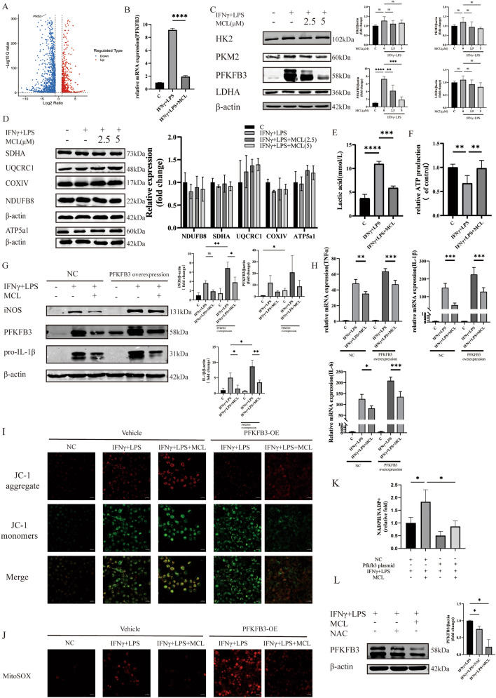
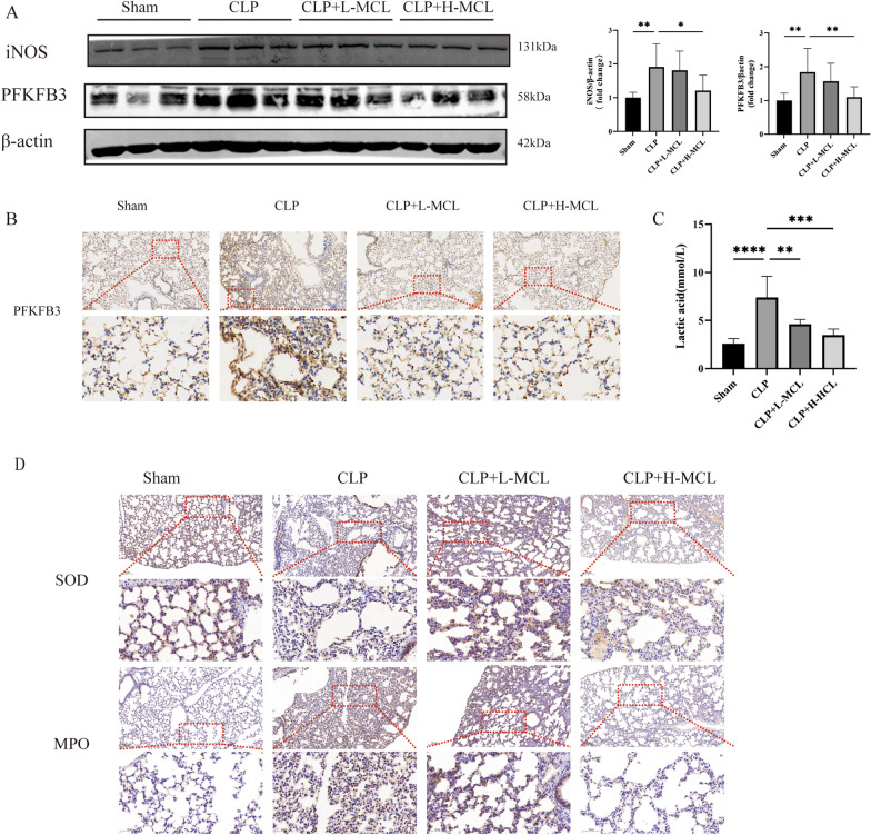
Methods: In vitro, lipopolysaccharides (LPS) and interferon-gamma (IFN-γ) were used to stimulate RAW264.7 cells and bone marrow-derived macrophages (BMDMs) to investigate the protective effect of MCL on sepsis-induced ALI. Cecal ligation and puncture (CLP) models were constructed in mice to induce ALI in vivo. The expression of inflammatory factors, macrophage polarization markers, and the glycolysis-related enzyme 6-phosphofructo-2-kinase/fructose-2,6-biphosphatase 3 (PFKFB3) were measured in vivo. Mitochondrial function, oxidative stress, and mitochondrial-related proteins were evaluated in vitro.

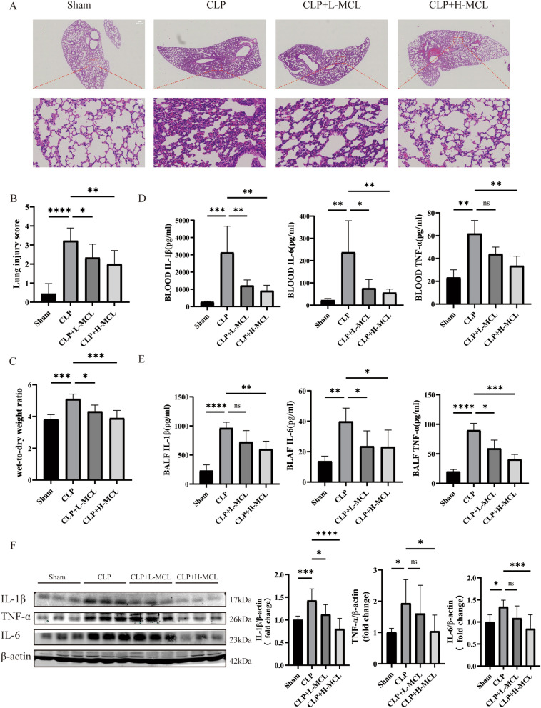
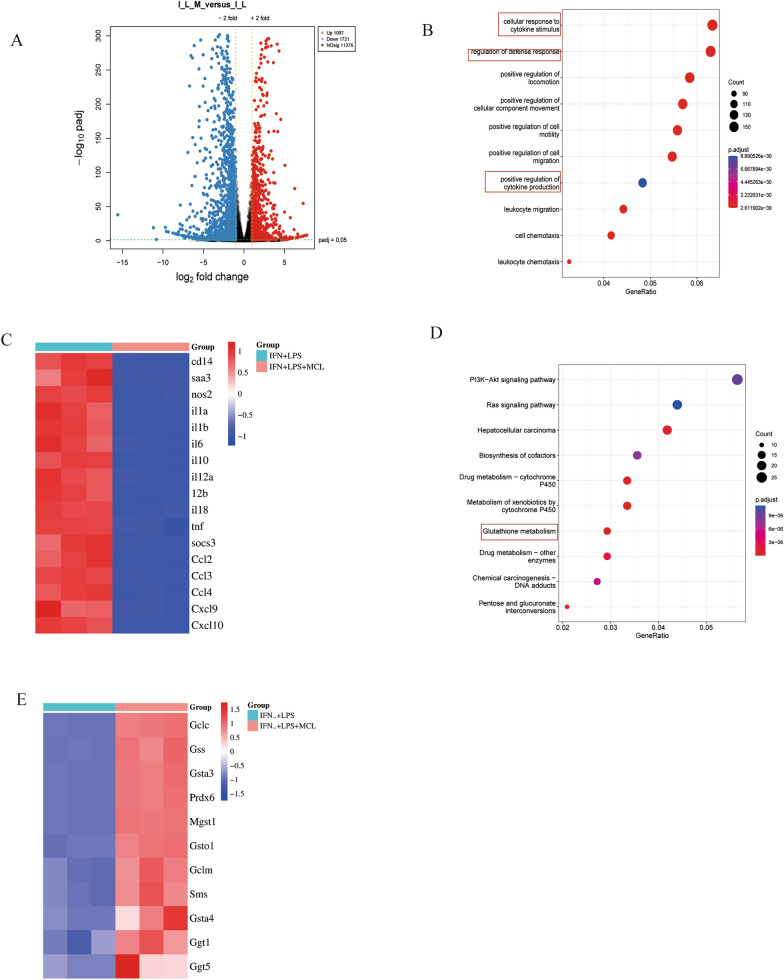
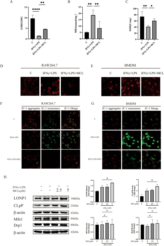
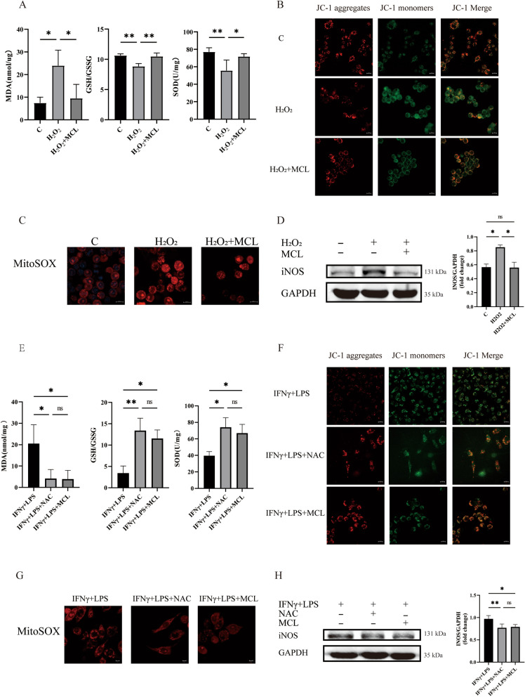
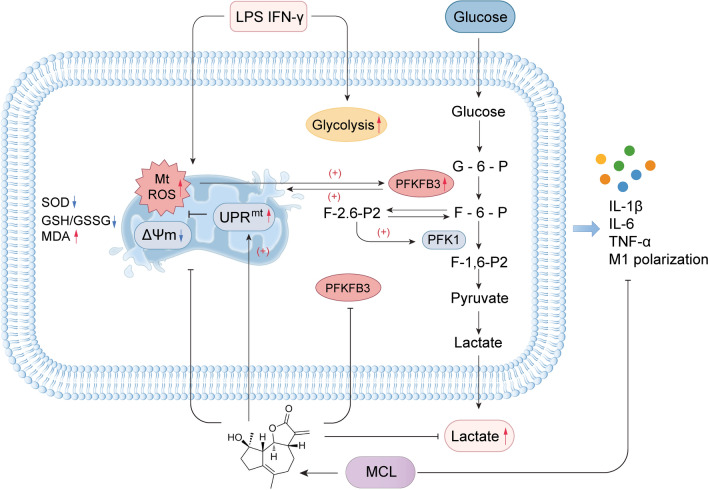
Results: MCL inhibited CLP-induced ALI, as evidenced by improvements in proinflammatory factor levels, lung wet/dry ratios, and histopathological findings. In vitro, MCL treatment significantly suppressed LPS + IFN-γ-induced M1-type polarization of RAW264.7 cells and BMDMs, as well as the production of inflammatory factors and oxidative stress. Mechanistic experiments revealed that MCL suppresses PFKFB3-driven glycolysis to reduce inflammation and activates the mitochondrial unfolded protein response (UPRmt) to alleviate mitochondrial stress. However, the therapeutic effect of MCL was diminished when PFKFB3 was overexpressed in cells.

Conclusion: This study is the first to demonstrate that MCL attenuates sepsis-induced ALI by reducing M1-type macrophage polarization. Its therapeutic effect is closely related to the suppression of oxidative stress and PFKFB3-driven glycolysis.

Keywords: Acute lung injury; Glycolysis; Macrophage polarization; Oxidative stress; PFKFB3.


Conflict of interest statement

Declarations. Ethics approval and consent to participate: All mice experiments were approved by the Southern Medical University Committee on Ethics of Animal Experiments (application NO: IACUC-LAC-20230705–001). Competing interests: The authors declare that they have no competing interests.

Figures

Fig. 1
Fig. 1
MCL attenuated sepsis-induced ALI in vivo. A Lung tissue was analyzed using H&E staining (upper panel: 40 × , scale bar = 500 μm; bottom panel: 400 × , scale bar = 50 μm). B The lung injury score of lung tissues. C Measurement of the W/D ratio in the lungs of mice. n = 5. D Inflammatory cytokines IL-1β, IL-6, and TNF-α production in blood. n = 5. E Inflammatory cytokines IL-1β, IL-6, and TNF-α production in BALF. n = 5. F Expression of IL-1β, IL-6, and TNF-α in lung tissue detected using WB. n = 3. Data are presented as mean ± SD. *p < 0.05, **p < 0.01, ***p < 0.001, ****p < 0.0001, ns = not significant, compared with the CLP group
Fig. 2
Fig. 2
MCL ameliorates M1-type macrophage polarization and the inflammatory response induced by LPS and IFN-γ. A MCL structure. B, C RAW264.7 cells and BMDMs were exposed to MCL for 24 h, and cell viability was determined by CCK8. D Protein expression of IL-1β, IL-6, and TNF-α in RAW264.7 cells. E Protein expression of IL-1β, IL-6, and TNF-α in BMDMs. F Levels of IL-1β, IL-6, and TNF-α in the supernatants of RAW264.7 cells (MCL: 10 μM). G Levels of IL-1β, IL-6, and TNF-α in the supernatants of BMDMs (MCL: 5 μM). H Protein expression of iNOS in RAW264.7 cells. I Protein expression of iNOS in BMDMs. Data are presented as mean ± SD. *p < 0.05, **p < 0.01, ***p < 0.001, ****p < 0.0001, ns = not significant, compared with the IFNγ + LPS group (n = 3)
Fig. 3
Fig. 3
Targets of MCL acting on BMDMs. A Volcano plot of upregulated and downregulated genes (I_L_M means IFNγ + LPS + MCL group, I_L means IFNγ + LPS group). B GO enrichment analysis for IFNγ + LPS + MCL versus IFNγ + LPS. C Heatmap of genes related to inflammation. D Top 10 KEGG pathways enriched in IFNγ + LPS + MCL versus IFNγ + LPS. E Heatmap of genes in the glutathione pathway
Fig. 4
Fig. 4
MCL attenuates mitochondrial oxidative stress in macrophages. A, C The GSH:GSSG ratio, SOD activity, and MDA production were measured in BMDMs. D, E Mitochondrial ROS production was measured by MitoSOX™ Red in RAW264.7 cells and BMDMs (scale bar = 10 μm). F, G Mitochondrial membrane potential was estimated using JC-1 in both RAW264.7 cells and BMDMs (scale bar = 10 μm). H Protein levels of LONP1, CLpP, Drp1, and MFN1 were estimated in BMDMs using WB. Data are presented as mean ± SD. *p < 0.05, **p < 0.01, ****p < 0.0001, compared with the IFNγ + LPS group (n = 3)
Fig. 5
Fig. 5
MCL regulates M1-type macrophage polarization by reducing mitochondrial oxidative stress. (A-D) BMDMs treated with H₂O₂ (100 μM). A The GSH:GSSG ratio, SOD activity, and MDA production. B Mitochondrial membrane potential was estimated using JC-1 (scale bar = 10 μm). C Mitochondrial ROS production was measured by MitoSOX™ Red (scale bar = 10 μm). D iNOS protein level was detected using WB. EH Pretreated BMDMs with NAC (2 mM) and MCL (5 μM). E The GSH:GSSG ratio, SOD activity, and MDA production. F Mitochondrial membrane potential was estimated using the JC-1 kit (scale bar = 10 μm). G Mitochondrial ROS production was measured by MitoSOX™ Red (scale bar = 10 μm). H iNOS protein level was analyzed using WB. Data are presented as mean ± SD. *p < 0.05, **p < 0.01, ns = not significant (n = 3)
Fig. 6
Fig. 6
MCL regulates PFKFB3-driven glycolysis. A Volcano plot showing PFKFB3 downregulation after MCL pretreatment (LPS + IFNγ + MCL group vs LPS + IFNγ group). B Relative mRNA expression of PFKFB3. C WB was used to quantify the protein levels of partial glycolysis-related proteins. D Levels of partial respiratory chain proteins were measured using WB. E Lactic acid levels in BMDM supernatants. F ATP levels. G iNOS and IL-1β expression after PFKFB3 overexpression. H IL-1β, IL-6, and TNF-α mRNA expression following PFKFB3 overexpression. I Mitochondrial membrane potential was estimated using the JC-1 kit (scale bar = 10 μm). J Mitochondrial ROS production was measured by MitoSOX™ Red (scale bar = 10 μm). K Change in the NADPH/NADP⁺ ratio after PFKFB3 overexpression. L PFKFB3 expression level after NAC pretreatment. Data are presented as mean ± SD. *p < 0.05, **p < 0.01, ***p < 0.001, ****p < 0.0001 (n = 3)
Fig. 7
Fig. 7
MCL protects mice from sepsis-induced ALI. A WB to assess iNOS and PFKFB3 levels in lung homogenates. B Expression of PFKFB3 was detected using immunohistochemistry (scale bar = 100 μm). C Lactic acid levels in mouse blood. D Expression of MPO and SOD was detected using immunohistochemistry (scale bar = 100 μm). Data are presented as mean ± SD. *p < 0.05, **p < 0.01, ***p < 0.001, ****p < 0.0001 (n = 3)
Fig. 8
Fig. 8
Micheliolide attenuates sepsis-induced ALI by suppressing oxidative stress and PFKFB3-driven glycolysis

References

    1. Kumar V. Pulmonary innate immune response determines the outcome of inflammation during pneumonia and sepsis-associated acute lung injury. Front Immunol. 2020;11:1722. - PMC - PubMed
    1. Cheng B, Xie G, Yao S, Wu X, Guo Q, Gu M, Fang Q, Xu Q, Wang D, Jin Y, et al. Epidemiology of severe sepsis in critically ill surgical patients in ten university hospitals in China. Crit Care Med. 2007;35:2538–46. - PubMed
    1. Zhou J, Qian C, Zhao M, Yu X, Kang Y, Ma X, Ai Y, Xu Y, Liu D, An Y, et al. Epidemiology and outcome of severe sepsis and septic shock in intensive care units in mainland China. PLoS ONE. 2014;9: e107181. - PMC - PubMed
    1. Sun JX, Xu XH, Jin L. Effects of metabolism on macrophage polarization under different disease backgrounds. Front Immunol. 2022;13: 880286. - PMC - PubMed
    1. He L, Jhong JH, Chen Q, Huang KY, Strittmatter K, Kreuzer J, DeRan M, Wu X, Lee TY, Slavov N, et al. Global characterization of macrophage polarization mechanisms and identification of M2-type polarization inhibitors. Cell Rep. 2021;37: 109955. - PMC - PubMed

MeSH terms

LinkOut - more resources