

Dental Calculus Formation Rate: The Role of Salivary Proteome and Metaproteome
- PMID: 39953744
- PMCID: PMC11949622
- DOI: 10.1111/jcpe.14142
Dental Calculus Formation Rate: The Role of Salivary Proteome and Metaproteome
Abstract
Background: Dental calculus accumulation varies across individuals. While various factors contribute to its accumulation, the role of salivary composition remains underexplored. This study aims to compare individuals suffering from rapid rates of dental calculus formation rates with those having slow formation rates in terms of salivary electrochemical properties as well as its proteomic, metaproteomic and elemental composition.
Methods: A total of 26 patients with a history of dental calculus were recruited. Saliva samples were collected and evaluated for electrochemical properties as well as elemental, proteomic and metaproteomic composition. Patients were provided scaling treatment to remove all calculus. Six months after the dental cleaning patients were re-assessed for the presence of dental calculus. Based on the dental calculus formation rate participants were categorised into slow (57.7%) and rapid calculus formers (42.3%) that were then assessed for differences in salivary composition.
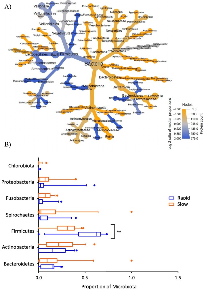
Results: Rapid calculus formers exhibited a more neutral zeta-potential and lower concentration of salivary calcium ions than their slow-forming counterparts. Proteomic analysis identified 895 proteins across all samples. Of these, 38 proteins were exclusive to the rapid formation group, while 24 proteins were specific to the slow group. The rapid group demonstrated augmented pathways related to cell binding (e.g., cytoskeletal regulation by Rho GTPase and integrin signalling), inflammatory mediation (e.g., chemokine and cytokine signalling) and neurodegenerative disorders (e.g., 5-Hydroxytryptamine degradation, Huntington's disease and Parkinson's disease) and significant enrichment in peptidase inhibitor activity. In contrast, the slow group demonstrated enrichment mainly in immune response. Metaproteomic analysis for salivary bacteria showed significant predominance of Streptococci in the rapid group and elevated levels of Rothia in the slow group.
Conclusion: The saliva of patients with rapid calculus formation rates differs from that of patients with slow rates of calculus formation in terms of electrochemical properties as well as proteomic, metaproteomic and elemental composition.
Keywords: dental calculus; human saliva; metaproteome; proteomics; salivary proteome; zeta potential.
© 2025 The Author(s). Journal of Clinical Periodontology published by John Wiley & Sons Ltd.
Conflict of interest statement
The authors declare no conflicts of interest.
Figures


References
-
- Afacan, B. , Öztürk V. Ö., Emingil G., Köse T., Mitsakakis K., and Bostanci N.. 2022. “Salivary Secretory Leukocyte Protease Inhibitor Levels in Patients With Stage 3 Grade C Periodontitis: A Comparative Cross‐Sectional Study.” Scientific Reports 12, no. 1: 21267. 10.1038/s41598-022-24295-2. - DOI - PMC - PubMed
Publication types
MeSH terms
Substances
Grants and funding
LinkOut - more resources
Full Text Sources
