. 2025 Feb 25;19(7):7005-7025.
doi: 10.1021/acsnano.4c14735. Epub 2025 Feb 15.

Multilayer Adjuvanted Influenza Protein Nanoparticles Improve Intranasal Delivery and Antigen-Specific Immunity

Affiliations

Multilayer Adjuvanted Influenza Protein Nanoparticles Improve Intranasal Delivery and Antigen-Specific Immunity

Jaeyoung Park et al. ACS Nano. .

Abstract

Intranasal vaccination is a desired route for protection against influenza viruses by mucosal and systemic immunity. However, the nasal mucosa impedes the intranasal delivery of vaccines. Here, we formulated layer-by-layer (LBL) influenza vaccine nanoparticles for effective intranasal delivery by coating them with alternating mucoadhesive cationic chitosan and muco-inert anionic CpG adjuvants. The nanoparticle cores were formed by desolvating influenza M2e antigen and coating it with hemagglutinin (HA) antigen via biotin-streptavidin conjugation. LBL modification promoted nasal delivery and interaction with the resident immune cells. Intranasal administration with LBL nanoparticles significantly improved cellular and humoral immune responses against HA and M2e including high IgA titers, a hallmark of potent mucosal immunity and persistence of immune responses. Distinct trends for antigen-specific immune responses were observed for different routes of vaccination. The enhanced immune responses conferred mice protection against the influenza challenge and prominently reduced viral titers, demonstrating the effectiveness of intranasal LBL vaccine nanoparticles.

Keywords: biodistribution; influenza vaccine; intranasal; layer-by-layer; mucosal immunology; nanoparticle; subunit vaccine.


Conflict of interest statement

The authors declare no competing financial interest.

Figures

Figure 1
Figure 1
In vivo biodistribution study. (a) IVIS spectrum images of mice administered IN with PBS, OVA NPs, PEG OVA NPs, and LBL OVA NPs 1 h after administration. (b) Profiles of radiant efficiency in mice over time. (c, d) Histology of NALT 24 h after nasal administration and lung stained with DAPI (blue) for cells, PE anti-CD19 for B cells (yellow), and FITC anti-CD11b for leukocytes (green) and biodistribution of PBS, OVA NPs (red), PEG OVA NPs (red), and LBL OVA NPs (red). (e) Uptake of OVA, PEG OVA, and LBL OVA NPs isolated from NALT by single cells, which were stained by anti-CD11b (biomarker for DCs) antibody or anti-CD19 (biomarker for B cells) antibody, and percent population of CD19+ cells isolated from NALT of mice treated with NPs. The brightness and contrast of histology images were uniformly adjusted to improve visualization of the red fluorescence of the NPs. The p values (n = 3) were determined by one-way ANOVA with Tukey’s post hoc multiple comparison analysis: ns for not significant, * for ≤0.05, ** for ≤0.01, *** for ≤0.001, and **** for ≤0.0001.
Figure 2
Figure 2
Characterization of LBL HA-4M2e NPs. (a) Diagram of HA-triGCN4 and 4M2e-tetraGCN4 proteins and their compositions. (b) Schematic illustration of LBL HA-4M2e NP synthesis. (c, d) Size distribution and hydrodynamic sizes (diameter) measured by dynamic light scattering (DLS). (e) Zeta potential measured by electrophoretic light scattering indicates the surface charges of NPs. (f, g) TEM images of HA-4M2e NPs and LBL HA-4M2e NPs. (h) Hemagglutination assay for the assessment of the functional structure of HA conjugated to 4M2e NPs. The data for nanoparticle size, PDI, and zeta potential were collected from three separate batches of NPs.
Figure 3
Figure 3
Systemic and local humoral immune responses against HA and 4M2e. (a) Different routes of vaccination: intramuscular priming and boost, intranasal priming and boost, and intramuscular priming followed by intranasal boost. (b) Schematic representation of the timeline for vaccination and collection of sera and BAL fluid. (c, d) Anti-HA (c) and anti-4M2e (d) IgG, IgG2a, and IgG1 titers in sera collected 49 days postpriming from mice immunized with HA + 4M2e + Adj, HA-4M2e NP, and LBL HA-4M2e NP. (e, f) Anti-HA (e) and anti-4M2e (f) IgA titers in BAL fluid collected 56 days postpriming from mice immunized. PBS-treated mice titers were below the limit of detection, so they are not shown. The p values (n = 5) were determined by two-way ANOVA with Tukey’s post hoc multiple comparison analysis: * for ≤0.05, ** for ≤0.01, *** for ≤0.001, and **** for ≤0.0001. ND represents not detectable values.
Figure 4
Figure 4
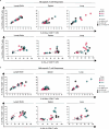
Antigen-specific cellular immune responses at 8 weeks post prime. (a) Percent populations of restimulated HA-specific IFN-γ+ CD8+ T cells, IFN-γ+ CD4+ T cells, and IL-4+ CD4+ T cells in the lymph node, spleen, and lung. (b) Percent populations of restimulated M2e-specific IFN-γ+ CD8+ T cells, IFN-γ+ CD4+ T cells, and IL-4+ CD4+ T cells. Comparison of the percentage of T-cell populations generally shows potent HA- and M2e-specific cellular immune responses induced by LBL HA-4M2e NPs especially when mice were administered via IN/IN and IM/IM or IM/IN routes, respectively. Bar graphs of the same data showing statistical analysis are shown in Figure S7.
Figure 5
Figure 5
Persistence of humoral immune responses against HA and 4M2e. (a) Timeline for vaccination and collection of sera, BAL fluid, and bone marrow. (b, c) Anti-HA (b) and anti-4M2e (c) IgG titers in sera collected on weeks 3, 7, 12, 16, and 20 postprime from mice immunized IM/IM and IN/IN with HA + 4M2e + Adj, HA-4M2e NP, and LBL HA-4M2e NP. (d, e) Anti-HA (d) and anti-4M2e (e) IgA titers in BAL fluid collected on 140 days postprime. (f–i) Quantification of HA- and 4M2e-specific IgG (f, g) and IgA (h, i) secreting long-lived bone marrow B cells after stimulating with R848/IL-2 (B-Poly-S reagent). PBS-treated mice titers were below the limit of detection and are not shown. ND stands for not detectable values. Comparisons across groups are only shown for week 20 time point. The p values (n = 5) were determined by two-way ANOVA with Tukey’s post hoc multiple comparison analysis: ns for not significant, * for ≤0.05, ** for ≤0.01, *** for ≤0.001, and **** for ≤0.0001.
Figure 6
Figure 6
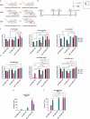
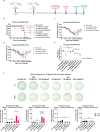
Immune protection conferred against challenges with H1N1 and pdm09 influenza A viruses in mice. (a) Timeline for intramuscular and intranasal vaccination and lethal virus challenge. (b–d) Survival rate (b) and body weight (c, d) of mice (n = 5) that were challenged with 4× LD50 H1N1 influenza A virus (A/California/04/2009) 6 weeks after the second vaccination. (e) Lung viral titers (n = 5) were measured from vaccinated mice after being challenged with 3× LD50 H1N1 influenza A virus 10.5 weeks after the second vaccination. The dashed line is the limit of detection. (f) M2e- and HA-specific IFN-γ (blue spots) and IL-2 (red spots) secreting splenocytes were quantified by T-ELISpot at 16 weeks after a booster. ND stands for not detectable values. Survival analysis was performed by Kaplan–Meier analysis with the log rank test: ns for not significant, * for ≤0.033, and ** for ≤0.002 compared to PBS control group. The p values (n = 5) were determined by two-way ANOVA with Tukey’s post hoc multiple comparison analysis: ns for not significant, * for ≤0.05, ** for ≤0.01, *** for ≤0.001, and **** for ≤0.0001.

References

    1. Vemula S. V.; Sayedahmed E. E.; Sambhara S.; Mittal S. K. Vaccine approaches conferring cross-protection against influenza viruses. Expert Rev. Vaccines 2017, 16 (11), 1141–1154. 10.1080/14760584.2017.1379396. - DOI - PMC - PubMed
    1. Harper S. A.; Fukuda K.; Uyeki T. M.; Cox N. J.; Bridges C. B. Prevention and control of influenza: Recommendations of the Advisory Committee on Immunization Practices (ACIP). Morb. Mortal. Wkly. Rep. 2005, 54 (Rr-8), 1–40. - PubMed
    1. Barría M. I.; Garrido J. L.; Stein C.; Scher E.; Ge Y.; Engel S. M.; Kraus T. A.; Banach D.; Moran T. M. Localized mucosal response to intranasal live attenuated influenza vaccine in adults. J. Infect Dis 2013, 207 (1), 115–124. 10.1093/infdis/jis641. - DOI - PMC - PubMed
    1. Gianchecchi E.; Manenti A.; Kistner O.; Trombetta C.; Manini I.; Montomoli E. How to assess the effectiveness of nasal influenza vaccines? Role and measurement of sIgA in mucosal secretions. Influenza and Other Respiratory Viruses 2019, 13 (5), 429–437. 10.1111/irv.12664. - DOI - PMC - PubMed
    1. Oh J. E.; Song E.; Moriyama M.; Wong P.; Zhang S.; Jiang R.; Strohmeier S.; Kleinstein S. H.; Krammer F.; Iwasaki A. Intranasal priming induces local lung-resident B cell populations that secrete protective mucosal antiviral IgA. Science Immunology 2021, 6 (66), eabj512910.1126/sciimmunol.abj5129. - DOI - PMC - PubMed

Publication types

MeSH terms