Published Erratum
. 2025 Feb 15;11(1):17.
doi: 10.1038/s41421-025-00773-5.

Author Correction: SGF29 nuclear condensates reinforce cellular aging

Affiliations
Published Erratum

Author Correction: SGF29 nuclear condensates reinforce cellular aging

Kaowen Yan et al. Cell Discov. .
No abstract available


Figures

Fig. 5
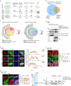
Fig. 5. Identification of SGF29 interacting proteins sensitive to condensate perturbation.
a Flow chart of the mass spectrometry strategy for the identification of EGFP-SGF29-WT (WT) and EGFP-SGF29-R207P (R207P) interacting proteins. Flag-EGFP was used as a control. b Shared and specific interacting proteins of EGFP-SGF29-WT (WT) and EGFP-SGF29-R207P (R207P) identified by mass spectrometry. c Chord diagrams showing the enriched pathways of shared interaction partners of EGFP-SGF29-WT (WT) and EGFP-SGF29-R207P (R207P) (left) and those of specific protein interaction partners of EGFP-SGF29-WT (right). d Co-IP analysis showing the interaction between indicated proteins and EGFP-SGF29-WT (WT) and EGFP-SGF29-R207P (R207P) in hMPCs. e Immunofluorescence staining of MED4 in hMPCs transduced with lentiviruses expressing either EGFP, EGFPSGF29-WT (WT) or EGFP-SGF29-R207P (R207P). Left, representative images. Scale bar, 10 μm. Right, quantification of the fluorescence intensity along the line embedded the image following the arrow direction. f Immunofluorescence staining of Pol II S2 and SGF29 in senescent hMPCs. Left, representative images. Scale bars, 10 μm and 5 μm (zoomed-in image). Right, quantification of the fluorescence intensity along the line embedded the image following the arrow direction. g Immunofluorescence staining of SP1 and SGF29 in senescent hMPCs. Left, representative images. Scale bars, 10 μm and 5 μm (zoomed-in image). Right, quantification of the fluorescence intensity along the line embedded the image following the arrow direction. h Coomassie blue staining of purified recombinant SGF29-C-D194A and SGF29-C-R207P after being resolved on SDS-PAGE. i Pelleting assay show that SGF29-C-D194A and SGF29-C-R207P interact with MED4 and SP1.

Erratum for

  • SGF29 nuclear condensates reinforce cellular aging.
    Yan K, Ji Q, Zhao D, Li M, Sun X, Wang Z, Liu X, Liu Z, Li H, Ding Y, Wang S, Belmonte JCI, Qu J, Zhang W, Liu GH. Yan K, et al. Cell Discov. 2023 Nov 7;9(1):110. doi: 10.1038/s41421-023-00602-7. Cell Discov. 2023. PMID: 37935676 Free PMC article.

References

    1. Yan, K. et al. SGF29 nuclear condensates reinforce cellular aging. Cell Discov.9, 110 (2023). - PMC - PubMed

Publication types

LinkOut - more resources