. 2025 Feb 15;16(1):1669.
doi: 10.1038/s41467-025-56601-7.

Discovery of selective low molecular weight interleukin-36 receptor antagonists by encoded library technologies

Affiliations

Discovery of selective low molecular weight interleukin-36 receptor antagonists by encoded library technologies

Juraj Velcicky et al. Nat Commun. .

Abstract

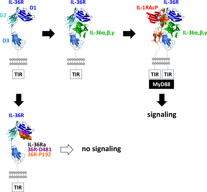
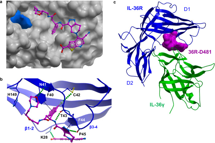
Interleukin-36 receptor (IL-36R), belonging to the IL-1 receptor family, is crucial for host defense and tissue repair. Targeting cytokine receptors with low molecular weight (LMW) compounds remains challenging due to their interaction with the large surface area of cytokine. In this study, two encoded library technologies are used to identify LMW molecules binding to IL-36R's extracellular domain. The mRNA-based display technique identifies 36R-P138, a macrocyclic peptide blocking IL-36R signaling. Importantly, its optimized analog (36R-P192) also effectively suppresses expression of marker genes induced by IL-36 in human skin biopsies. DNA encoded libraries (DEL) screening delivers 36R-D481, a high affinity LMW IL-36R binder, effectively inhibiting IL-36 signaling. X-ray crystallography analysis reveals that both the cyclic peptide and DEL-compound bind to the IL-36R's D1 domain, potentially disrupting IL-36 cytokine binding. This study demonstrates that it is possible to target a cytokine receptor within the IL-1 receptor family using a small molecule ( < 1000 Da).


Conflict of interest statement

Competing interests: All authors are or were at the time of their involvement with the research employees of Novartis BioMedical Research and may hold stocks in Novartis. There are no more competing interests.

Figures

Fig. 1
Fig. 1. Formation of the ternary IL36/IL-36R/IL-1RAcP complex.
Signaling through IL-36R relies on the formation of the IL-36 ternary complex, which occurs in two steps. In the first step, one of the agonistic IL-36 cytokines binds to IL-36R. The formed binary complex then associates with IL-1RAcP, resulting in the proximity of the TIR domains on both IL-36R and IL-1RAcP that is essential for downstream signaling through MyD88. Binding of IL-36 agonists and signaling through IL-36R is blocked by the natural antagonist (IL-36Ra) or by IL-36 inhibitors described in this paper.
Fig. 2
Fig. 2. mRNA display screen identified a high affinity binder to IL-36R.
a Illustrative mRNA display selection cycle used to identify macrocyclic peptides against IL-36R. Selections were performed with and without addition of the spesolimab-Fab. b Chemical structures of peptides 36R-P138 and 36R-P192, amino acid replacements shown in red. c Affinities of 36R-138 and 36R-P192 to IL-36R measured by SPR. d Single cycle kinetics shown for 36R-P192/IL-36R D1-D3 with 36R-P192 concentrations from 0.4 to 100 nM; experimental data in black, fit in red. e SPR measurements of IL-36α/IL-36R D1-D3 with IL-36α concentrations from 0.02 to 2.5 µM (left panel), binding of IL-36α at 0.6 µM to IL-36R D1-D3 in the presence of 36R-P192 (middle panel), quantification of competition of IL-36α binding IL36R D1-D3 at 0.6 µM with 36R-P192 (right panel).
Fig. 3
Fig. 3. Peptides 36R-P138 and 36R-P192 inhibit IL-36R signaling in cellular assays and human skin biopsies.
a Left panel, HekBlue_IL-36R cells had been stimulated with either 0.03 ng/mL IL-36α or 5 ng/mL IL-1β in the absence or presence of 36R-P138 and 36R-P192, respectively and SEAP was measured after 6 h; right panel, A431 cells had been stimulated with either 10 ng/mL IL-36α or 5 ng/mL IL-1β in the absence or presence of 36R-P138 and 36R-P192, respectively and released IL-8 was measured in cell supernatants after 18 h of incubation. Representative experiment is shown and mean IC50’s with stdv from three independent experiments are indicated in the boxes. b Effect of 36R-P192 in dose response on SEAP expression of HekBlue_IL-36R cells upon stimulation with various concentrations of IL-36α (upper panel), IL-36β (middle panel) and IL-36γ (lower panel). Experiment was done in duplicates and a representative experiment is shown. c Skin biopsies from 4-6 different donors had been pre-incubated for 30 min with either 200 nM spesolimab or various concentrations of 36R-P192 and stimulated with 50 ng/mL IL-36α for 24 h; mRNA was extracted, and marker gene expression was quantified by qPCR and calculated as fold induction compared to unstimulated controls. Statistical analysis was conducted using ordinary one-way ANOVA (multiple comparison) and error bars indicate the standard deviation; * p < 0.05, ** p < 0.01, *** p < 0.001, **** p < 0.0001. Source data are provided as a Source Data file.
Fig. 4
Fig. 4. 36R-D481 binds to IL-36R and inhibits IL-36R signaling.
a Chemical structure of 36R-D481 and SPR measurements of 36R-D481 binding to IL-36R; calculated Kd’s for binding either to IL-36R D1-D2 or IL-36R D1-D3 are indicated in the table. b HekBlue_IL-36 R cells had been stimulated with either 0.01 ng/mL IL-36α, 0.01 ng/mL IL-36γ, 0.003 ng/mL IL-36β or 5 ng/mL IL-1β in the absence or presence of 36R-D481 and SEAP was measured after 6 h; representative experiment is shown and mean IC50’s with stdv from three independent experiments are shown in the table. c Effect of 36R-D481 measured in dose response on HekBlue_IL-36R cells in the presence and of various concentrations of IL-36α. Experiment was done in duplicates and a representative experiment is shown. Source data are provided as a Source Data file.
Fig. 5
Fig. 5. Crystal structure of 36R-P192 bound to IL-36R D1-D2/spesolimab-Fab complex.
a 36R-P192 (moccasin) shown as solvent accessible surface bound to IL-36R D1-D2 (blue ribbon) and spesolimab-Fab (ribbon; HC in magenta, LC in pink) complex (PDB: 9ETH). b 36R-P192 (mocassin sticks) binding to IL-36R D1-D2 shown as solvent accessible surface. c Detailed intramolecular interactions within the cyclic peptide structure of 36R-P192. d, e Interactions of 36R-P192 with IL-36R D1-D2. f IL-36R D1-D2 (blue ribbon) with 36R-P192 (moccasin, skin representation) and IL-36γ (green ribbon) (PDB: 4IZE) superimposed to ternary IL1β/IL-1R1/IL-1RAcP (PDB: 4DEP, not shown, only used for the overlay). The modelled structure demonstrates possible clash between 36R-P192 and IL-36γ, thus competing for the same binding site on the concave D1-D2 site of the IL-36 receptor.
Fig. 6
Fig. 6. Crystal structure of 36R-D481 bound to IL-36R D1-D2/spesolimab-Fab complex.
a 36R-D481 (magenta) bound to IL-36R D1-D2 (water accessible surface shown in skin representation, PDB: 9ETI). Pocket between D1 and D2 shown in blue. b Key interactions between 36R-D481 bound to IL36R (blue). c Possible interaction of IL-36R D1-D2 (binding 36R-D481) with IL-36γ (green, PDB: 4IZE) modelled by superimposition of IL36R D1-D2 with the D1-D2 domains of IL-1R1 and IL-36γ superimposed with IL1β in the ternary IL1β/IL-1R1/IL-1RAcP complex (PDB: 4DEP). The modelled complex structure demonstrates a possible clash between 36R-D481 (magenta) and IL-36γ (green).

References

    1. Gabay, C. & Towne, J. E. Regulation and function of interleukin-36 cytokines in homeostasis and pathological conditions. J. Leukoc. Biol.97, 645–652 (2015). - PubMed
    1. Bassoy, E. Y., Towne, J. E. & Gabay, C. Regulation and function of interleukin-36 cytokines. Immunol. Rev.281, 169–178 (2018). - PubMed
    1. Xu, P., Guan, H., Xiao, W. & Sun, L. The role of IL-36 subfamily in intestinal disease. Biochem Soc. Trans.50, 223–230 (2022). - PubMed
    1. Buhl, A. L. & Wenzel, J. Interleukin-36 in Infectious and Inflammatory Skin Diseases. Front Immunol.10, 1162 (2019). - PMC - PubMed
    1. Manzanares-Meza, L. D., Valle-Rios, R. & Medina-Contreras, O. Interleukin-1 Receptor-Like 2: One Receptor, Three Agonists, and Many Implications. J. Interferon Cytokine Res42, 49–61 (2022). - PubMed

MeSH terms

LinkOut - more resources