. 2025 Feb 15;22(1):41.
doi: 10.1186/s12974-025-03345-2.

Human breast milk-derived exosomes attenuate lipopolysaccharide-induced activation in microglia

Affiliations

Human breast milk-derived exosomes attenuate lipopolysaccharide-induced activation in microglia

Oluwatomi Akinduro et al. J Neuroinflammation. .

Abstract

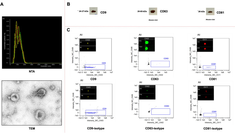
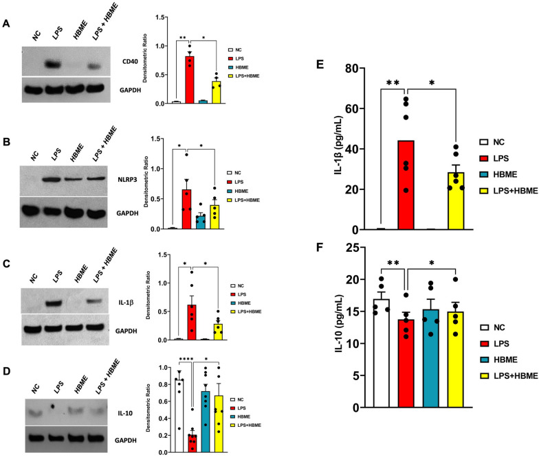
Microglia mediate the immune response in the central nervous system to many insults, including lipopolysaccharide (LPS), a bacterial endotoxin that initiates neuroinflammation in the neonatal population, especially preterm infants. The synthesis of the proinflammatory proteins CD40 and NLRP3 depends on the canonical NF-κB cascade as the genes encoding CD40 and NLRP3 are transcribed by the phosphorylated NF-κB p50/p65 heterodimer in LPS-induced microglia. Exosomes, which are nanosized vesicles (40-150 nm) involved in intercellular communication, are implicated in many pathophysiological processes. Human breast milk, which is rich in exosomes, plays a vital role in neonatal immune system maturation and adaptation. Activated microglia may cause brain-associated injuries or disorders; therefore, we hypothesize that human breast milk-derived exosomes (HBME) attenuate LPS-induced activation of CD40 and NLRP3 by decreasing p38 MAPK and NF-κB p50/p65 activation/phosphorylation downstream of TLR4 in murine microglia (BV2). Human microglia (HMC3) showed a significant decrease in p65 phosphorylation. We isolated purified HBME and characterized them using nanoparticle tracking analysis, transmission electron microscopy, fluorescence-activated cell sorting, and western blots. Analysis of microglia exposed to LPS and HBME indicated that HBME modulated the expression of signaling molecules in the canonical NF-κB pathway, including MyD88, IκBα, p38 MAPK, NF-κB p65, and their products CD40, NLRP3, and cytokines IL-1β and IL-10. Thus, HBMEs have great potential for attenuating the microglial response to LPS.

Keywords: BV2; Breast milk; CD40; Exosomes; HMC3; IL-10; IL-1β; Lipopolysaccharide; Microglia; NFκB; NLRP3; Neonatal neuroinflammation.


Conflict of interest statement

Declarations. Ethics approval and consent to participate: Ethics approval and consent to participate were not needed. BV2 cells were a generous gift from Dr. Harald Neumann at the University of Bonn LIFE and Brain Center in Bonn, Germany. Consent for publication: Not applicable. Competing interests: The authors declare no competing interests.

Figures

Fig. 1
Fig. 1
HBME inhibit LPS activated canonical NF-kB pathway in microglia and its effectors. HBME seems to enact inhibitory effect either upstream of the TLR4 signaling pathway or directly on different molecules within the cascade. LPS activates the TLR4 receptor causing the activation of two proinflammatory transcription factors, p38 MAPK and the NF-kB p50/p65 heterodimer, ultimately leading to the increased production of key promotors of the microglial immune response. TLR4 relies on the subsequent activation of its adapter protein, MyD88 to potentiate downstream protein activation. The phosphorylated p38 MAPK translocates directly to the nucleus to produce the proinflammatory mediators IL-1b and NO, and the anti-inflammatory cytokine IL-10 through downstream mechanisms. Separately, p38 MAPK further potentiates the activation of NF-kB. The literature suggests that p38 phosphorylates IkBa just as the IKK trimer complex does, thus freeing NF-kB to phosphorylate and translocate to the nucleus where it produces key proinflammatory mediators. CD40 will be incorporated into the plasma membrane to recognize its ligand and further activate microglia. Pro-IL-1b must be cleaved into its active form via the caspase-1 protease found within the NLPR3 inflammasome’s multi-protein structure. For NLRP3 to become active and integrated into its inflammasome, a secondary PAMP or DAMP such as the HMGB1 protein must activate it. IL-1b and NO, along with several other proinflammatory cytokines and chemokines, are released from microglia propagating a neuroinflammatory response. Due to these mounting effects, microglial activation can lead to an exaggerated inflammatory response and subsequent CNS toxicity. HBME: Human breast milk derived exosomes; LPS: Lipopolysaccharide; TLR4: Toll-Like Receptor 4; MAPK: Mitogen-activated protein kinases; NF-kB: Nuclear factor kappa B; MyD88: Myeloid differentiation primary response 88; IL-1b: interleukin one beta; NO: nitric oxide; IL-10: interleukin 10; IkBa: Nuclear factor of kappa light polypeptide gene enhancer in B-cells inhibitor alpha; IKK: Inhibitor of nuclear factor-κB kinase; CD40: Cluster of differentiation 40; NLRP3: nucleotide-binding oligomerization domain-like receptor protein 3; PAMP: Pathogen-associated molecular patterns; DAMP: Damage-associated molecular patterns; HMGB1: High mobility group box1
Fig. 2
Fig. 2
Characterization of Human Breast Milk-derived Exosomes. A Exosome size, number, and morphology were evaluated using NTA and TEM and were found to be in the expected size range and of the expected morphology. Exosome expression of the tetraspanin molecules CD9, CD63, and CD81 were verified using B WB analysis to demonstrate the presence of these exosome-specific markers in exosome protein lysate and C FACS analysis to demonstrate their presence on the surface of live, intact exosomes. Each tetraspanin was compared to a IgG isotype control to account for nonspecific binding. n ≥ 3. NTA: Nanosight tracking analysis; TEM: transmission electron microscopy; WB: western blot; FACS: fluorescence-activated cell sorting
Fig. 3
Fig. 3
Effects of HBME on cell survival and CD40 expression in LPS-induced BV2 microglia. CD40 expression was determined in BV2 microglia treated with PBS (NC), LPS (1 µg/mL), and/or HBME (10 µg/mL) from a single breast milk sample for 24 h. A Survival of BV2 microglia cells was measured with trypan blue using a Countess 2 automated cell counter (n = 12) B, C Differential expression analysis for LPS-treated or LPS- and HBME-treated microglia. RNA-seq data were analyzed by edgeR (threshold was set as fold change > 1.5, p < 0.05). D 14 of the analyzed genes that are important in LPS-induced inflammatory processes were displayed on a heatmap for more direct comparison E RT-qPCR of gene encoding CD40 in multiple experiments (n = 3). F WB image for expression of CD40 with densitometry (Image J) analysis for each sample (n = 7). Quantified densitometric ratios normalized to GAPDH. Bars represent the mean ± SEM, n ≥ 3/group, *p < 0.05, **p < 0.01, ***p < 0.001, ****p < 0.0001 by ANOVA followed by Fisher’s least significant difference post-hoc multiple comparison test. HBME, human breast milk-derived exosomes
Fig. 4
Fig. 4
Effects of HBME on the TLR4/NF-kB signaling pathways in LPS-induced BV2 microglia. Expression of proteins in the NF-kB pathway affected by LPS stimulation of microglia was determined by WB analysis. Cells were treated with PBS (NC), LPS (1 µg/mL), and/or HBME (10 µg/mL) for 15 min, 1 h, or 24 h. Quantified densitometric ratios normalized to GAPDH (A, B) or total p38 (C)/total NFkB p65 (D). Representative WB images of A MyD88, B IkBa, C MAP kinase p38, and D NF-kB p65 after exposure to experimental conditions are disclosed. Densitometry (Image J) analysis for each sample. Bars represent mean ± SEM, n = 3, *p < 0.05, **p < 0.01, ***p < 0.001, ****p < 0.0001 by one-way ANOVA followed by Fisher’s least significant difference post-hoc multiple comparison test
Fig. 5
Fig. 5
Effects of HBME on inflammatory markers in LPS-induced BV2 microglia. Expression of NF-kB pathway downstream effectors of inflammation by LPS stimulation of microglia was determined by WB analysis and ELISA. Cells were treated as previously described for 24 h. Quantified densitometric values were graphed and representative WB images were disclosed for A CD40 (n = 4), B NLRP3 (n = 5), C IL-1b (n = 6), and D IL-10 (n = 8). Densitometric (Image J) analysis for each sample. ELISA analysis represented for E IL-1b (n = 6) and F IL-10 (n = 5). Bars represent mean ± SEM, n ≥ 3, *p < 0.05, **p < 0.01, ***p < 0.001, ****p < 0.0001 by one-way ANOVA followed by Fisher’s least significant difference post-hoc multiple comparison test
Fig. 6
Fig. 6
HBME inhibit the proinflammatory response and morphological changes of LPS-induced microglia. Immunofluorescence staining on microglial cells demonstrates HBME-mediated reduction in morphological changes and proinflammatory marker expression reflective of LPS-induced microglial activation. A BV2 microglia were treated with PBS (NC), LPS (1 µg/mL), and/or HBME (5 µg/mL) for 24 h and probed for CD40, phalloidin (F-actin), and DAPI. B HMC3 microglia were treated with PBS (NC), LPS (1 µg/mL), and/or HBME (10 µg/mL) for 4 h and probed for Iba-1, phalloidin (F-actin), and DAPI
Fig. 7
Fig. 7
Effects of HBME on intracellular signaling and cytokine secretion from LPS-induced HMC3 microglia. Expression of proteins in the NF-kB pathway affected by LPS and/or HBME stimulation of microglia was determined by WB and ELISA analysis. Cells were treated with PBS (NC), LPS (1 µg/mL), and/or HBME (10 µg/mL) for 1 h. A Representative WB image and the quantified densitometric ratios normalized to GAPDH of NFkB p65 are shown (n = 6). B ELISA analysis of IL-1b secretion from cells into the media represented (n = 3). Bars represent mean ± SEM, n ≥ 3, *p < 0.05, **p < 0.01, ***p < 0.001, ****p < 0.0001 by one-way ANOVA followed by Fisher’s least significant difference post-hoc multiple comparison test

References

    1. Zhou Q, Guo D, Li X, Wang Y, Ye X, Xue S, et al. Anti-inflammatory effects of vinpocetine in LPS-stimulated microglia via activation of AMPK. An Acad Bras Ciênc. 2020;92(4): e20200241. - PubMed
    1. Sousa NA, Oliveira GAL, De Oliveira AP, Lopes ALF, Iles B, Nogueira KM, et al. Novel ocellatin peptides mitigate LPS-induced ROS formation and NF-kB activation in microglia and hippocampal neurons. Sci Rep. 2020;10(1):2696. - PMC - PubMed
    1. Li B, Dasgupta C, Huang L, Meng X, Zhang L. MiRNA-210 induces microglial activation and regulates microglia-mediated neuroinflammation in neonatal hypoxic-ischemic encephalopathy. Cell Mol Immunol. 2020;17(9):976–91. - PMC - PubMed
    1. Shemer A, Scheyltjens I, Frumer GR, Kim J-S, Grozovski J, Ayanaw S, et al. Interleukin-10 prevents pathological microglia hyperactivation following peripheral endotoxin challenge. Immunity. 2020;53(5):1033-49.e7. - PubMed
    1. Dean JM, van de Looij Y, Sizonenko SV, Lodygensky GA, Lazeyras F, Bolouri H, et al. Delayed cortical impairment following lipopolysaccharide exposure in preterm fetal sheep. Ann Neurol. 2011;70(5):846–56. - PubMed

Substances

LinkOut - more resources