Review
. 2025 Feb 15;22(1):39.
doi: 10.1186/s12974-025-03360-3.

Innate immune sensors and regulators at the blood brain barrier: focus on toll-like receptors and inflammasomes as mediators of neuro-immune crosstalk and inflammation

Affiliations
Review

Innate immune sensors and regulators at the blood brain barrier: focus on toll-like receptors and inflammasomes as mediators of neuro-immune crosstalk and inflammation

Cigdem Acioglu et al. J Neuroinflammation. .

Abstract

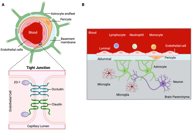
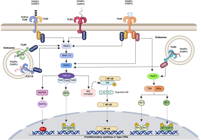
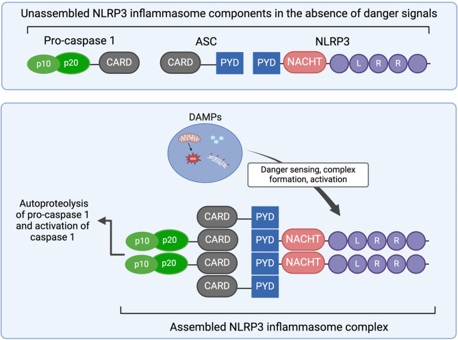
Cerebral endothelial cells (CEC) that form the brain capillaries are the principal constituents of the blood brain barrier (BBB), the main active interface between the blood and the brain which plays a protective role by restricting the infiltration of pathogens, harmful substances and immune cells into the brain while allowing the entry of essential nutrients. Aberrant CEC function often leads to increased permeability of the BBB altering the bidirectional communication between the brain and the bloodstream and facilitating the extravasation of immune cells into the brain. In addition to their role as essential gatekeepers of the BBB, CEC exhibit immune cell properties as they can receive and transmit signals between the blood and the brain partly via release of inflammatory effectors in pathological conditions. Cerebral endothelial cells express innate immune receptors, including toll like receptors (TLRs) and inflammasomes which are the first sensors of exogenous or endogenous dangers and initiators of immune and inflammatory responses which drive neural dysfunction and degeneration. Accumulating evidence indicates that activation of TLRs and inflammasomes in CEC compromises BBB integrity, promotes aberrant neuroimmune interactions and modulates both systemic and neuroinflammation, common pathological features of neurodegenerative and psychiatric diseases and central nervous system (CNS) infections and injuries. The goal of the present review is to provide an overview of the pivotal roles played by TLRs and inflammasomes in CEC function and discuss the molecular and cellular mechanisms by which they contribute to BBB disruption and neuroinflammation especially in the context of traumatic and ischemic brain injuries and brain infections. We will especially focus on the most recent advances and literature reports in the field to highlight the knowledge gaps. We will discuss future research directions that can advance our understanding of the central contribution of innate immune receptors to CEC and BBB dysfunction and the potential of innate immune receptors at the BBB as promising therapeutic targets in a wide variety of pathological conditions of the brain.

Keywords: Blood brain barrier; Brain injury; COVID-19.; Endothelial cells; Infection; Inflammasome; Neurovascular unit; Pericytes; Stroke; Toll like receptors.


Conflict of interest statement

Declarations. Ethics approval and Consent for Participation: Not applicable. Consent for publication: Not applicable. Competing interests: The authors declare no competing interests.

Figures

Fig. 1
Fig. 1
Cells of the blood brain barrier (BBB). (A) The principal cells of the BBB are cerebral endothelial cells (CEC) which form the capillaries of the central nervous system. Pericytes embedded in the basement membrane and astrocytes which send endfeet are additional components of the BBB. (B) Astrocytes also interact with neurons and microglia and mediate endothelial cell-neuron and endothelial cell-microglia communication. Created with BioRender.com
Fig. 2
Fig. 2
Toll-like receptor signaling pathways. TLR2, TLR4, and TLR5 are cell surface receptors whereas TLR3 and TLR7-9 are intracellular receptors. Most TLRs utilize the MyD88-dependent signaling pathway, except for TLR3, which signals through TRIF. Ligand binding to TLRs triggers the formation of homodimers. TLR2 forms heterodimers with TLR1 or TLR6. TLR2 and TLR4 interact with MyD88 via the bridging adaptor TIRAP. TLR4 also signals via the TRIF pathway and utilizes the bridging adaptor TRAM. Activation of the MyD88 signaling pathway leads to the activation of MAPKs resulting in AP-1 dependent transcription. Activation of IKK complex causes the translocation of NF-κB to the nucleus and initiation of NF-κB-mediated transcription. Translocation of IRF5 to the nucleus promotes IRF5-dependent transcription. Activation of the TRIF pathway induces either IRF3- and IRF7-dependent transcription, or AP-1, NF-κB and IRF5-dependent transcription through TRAF6. This leads to pro-inflammatory cytokine and Type 1 IFNs expression and release. AP-1, activator protein 1; IFN, interferon; IRAK, interleukin-1 receptor-associated kinase; IKK, IκB kinase; IRF, interferon regulatory factor; MyD88, myeloid differentiation primary response 88; MAPK, mitogen-activated protein kinase; NF-κB, nuclear factor kappa B; TLR, toll-like receptor; TIRAP, toll/interleukin-1 (IL-1) receptor (TIR) domain containing adaptor protein; TRAM, toll/IL-1R domain-containing adaptor-inducing IFN-β-related adaptor molecule; TRIF, Toll/IL-1R domain-containing adaptor-inducing IFN-β. Created with BioRender.com
Fig. 3
Fig. 3
Assembly of the NLRP3 inflammasome. Components of the NLRP3 inflammasome (upper panel). The components, pro-caspase-1, ASC and NLRP3 are not assembled in homeostatic conditions. NLRP3 contains a C-terminal LRR domain, a central NACHT domain, and N-terminal PYD. Upon danger sensing by NLRP3, complex assembly is initiated; NLRP3 oligomers are formed and recruit the adaptor protein ASC via PYD-PYD interactions whereas ASC interacts with pro-caspase-1 through CARD-CARD (lower panel). Following assembly, autoproteolysis of pro-caspase 1 leads to caspase-1 activation. ASC, apoptosis-associated speck-like protein containing a CARD (ASC); CARD, caspase activation and recruitment domain; DAMP, danger associated molecular pattern; LRR, leucine rich repeat; NLR, nucleotide binding and oligomerization domain (NOD)-like receptors. Created with BioRender.com
Fig. 4
Fig. 4
Activation of NLRP3 inflammasome. Activation of NLRP3 inflammasome involves two steps. The first step, priming (left panel) is triggered by cytokines or PAMPs/DAMPs acting through TLRs and leads to pro-IL-1β and pro-IL-18 production. The second step (right panel) is NLRP3 inflammasome assembly and activation, which is triggered by several signals, including K+ efflux, Ca2+ influx, extracellular ATP, lysosomal cathepsin and ROS. Increased intracellular Ca2+ due to ER stress causes mitochondrial damage and ROS release whereas ruptured lysosomes release cathepsin. This triggers the assembly and activation of NLRP3 inflammasome. Activation of caspase-1 results in the proteolytic cleavage of pro-IL-1β and pro-IL-18 and secretion of active IL-1β and IL-18. In addition, NLRP3 inflammasome activation results in cleavage of GSDMD. The N-terminal fragment (N-GSDMD) generated is incorporated into the cell membrane and forms pores that mediate pyroptosis. ASC, apoptosis-associated speck-like protein containing a CARD; CARD9, caspase recruitment domain-containing protein 9; DAMP, danger associated molecular pattern; GSDMD, Gasdermin D; IL-1β, interleukin 1β; IL-1R, interleukin receptor 1; NF-κB, nuclear factor kappa B; NLRP3, NACHT, LRR- and pyrin domains-containing protein 3; P2X, purinoceptor 7; PAMP, pathogen associated molecular pattern; ROS, reactive oxygen species; TLR, toll-like receptor; TNF, tumor necrosis factor; TNFR, TNF receptor. Created with BioRender.com
Fig. 5
Fig. 5
Non-canonical inflammasome activation. Sensing of intracellular LPS by pro-caspase-11 (or pro-caspase-4/5) leads to oligomerization and activation of the non-canonical inflammasome resulting in activation of caspase 11. Activated caspase-11 cleaves pore forming GSDMD, leading to pyroptosis. Caspase 11 activation also results in NLRP3 inflammasome activation, which, in turn, leads to IL-1β production and release. GSDMD, Gasdermin D; IL-1β, Interleukin β; LPS, Lipopolysaccharide; Created with BioRender.com

References

    1. Chen T, Dai Y, Hu C, Lin Z, Wang S, Yang J, Zeng L, Li S, Li W. Cellular and molecular mechanisms of the blood-brain barrier dysfunction in neurodegenerative diseases. Fluids Barriers CNS. 2024;21:60. - PMC - PubMed
    1. Lizano P, Pong S, Santarriaga S, Bannai D, Karmacharya R. Brain microvascular endothelial cells and blood-brain barrier dysfunction in psychotic disorders. Mol Psychiatry. 2023;28:3698–708. - PubMed
    1. Segarra M, Aburto MR, Acker-Palmer A. Blood-brain barrier dynamics to maintain brain homeostasis. Trends Neurosci. 2021;44:393–405. - PubMed
    1. Yuan Y, Sun J, Dong Q, Cui M. Blood-brain barrier endothelial cells in neurodegenerative diseases: signals from the barrier. Front Neurosci. 2023;17:1047778. - PMC - PubMed
    1. Girolamo F, Errede M, Bizzoca A, Virgintino D, Ribatti D. Central nervous system pericytes contribute to health and disease. Cells 2022, 11. - PMC - PubMed

MeSH terms

LinkOut - more resources