. 2025 Feb 16;16(1):1677.
doi: 10.1038/s41467-025-57001-7.

Tissue factor-dependent colitogenic CD4+ T cell thrombogenicity is regulated by activated protein C signalling

Affiliations

Tissue factor-dependent colitogenic CD4+ T cell thrombogenicity is regulated by activated protein C signalling

Gemma Leon et al. Nat Commun. .

Abstract

Patients with inflammatory bowel disease (IBD) have an increased risk of venous thromboembolism (VTE), but the underlying mechanistic basis remains poorly defined. Here, we find that colitogenic CD4+ T cells express tissue factor (TF) and promote rapid TF-dependent plasma thrombin generation. TF+CD3+CD4+ T cells are present in both the colons of mice with experimental colitis and blood and colonic tissue from patients with IBD. Expression of genes involved in regulating coagulation, including Protein C (PC; encoded by PROC) and its receptor (PROCR), are dysregulated in IBD patient gut biopsy tissues. Moreover, activated PC signalling reduces the procoagulant activity mediated by TF+CD4+ T cells. Our data thus identify TF-induced, colitogenic T cell-mediated thrombogenicity, and also demonstrate a new function for activated PC signalling in regulating T cell thrombo-inflammatory activity.


Conflict of interest statement

Competing interests: The authors declare no competing interests.

Figures
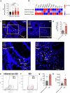
Fig. 1
Fig. 1. TF expression is upregulated during IBD.
a Expression of TF from RNA-seq data of ileal biopsies from Crohn’s Disease (CD); illeal (iCD, n = 162), colonic (cCD, n = 56), Ulcerative Colitis (UC, n = 62), and query IBD healthy control cohort (CTRL, n = 42) patients in the RISK study (GEO ID GSE57945). b Expression of coagulation and inflammation-associated genes from RNA sequencing of paediatric CD (n = 11), UC (n = 7), and inflamed query non-IBD biopsies (n = 12). c TF, CD3 and CD4 co-staining and d the average number of TF+ CD3+ CD4+ T cells per field in colonic biopsies from paediatric IBD patients (IBD, n = 4, non-IBD, n = 3). eh Lamina propria cells were isolated from colonic biopsies from paediatric IBD patients (n = 5) and inflamed query non-IBD biopsies (n = 2). eg The percentage of TF expressing CD3+ CD4+ TF+ T cells and h their cell surface TF expression was measured by flow cytometry and compared. Student’s t-test (two-tailed) (d, h) or Mann–Whitney U Test (a, g) was used to determine statistical significance from a 322 biological donors (iCD, n = 162, cCD, n = 56, UC, n = 62, control non-IBD group, n = 42), d 7 biological donors (n = 4 IBD, n = 3 inflamed non-IBD), g, h 7 biological donors (n = 5 IBD, n = 2 inflamed non-IBD) and expressed as mean ± s.e.m. Source data are provided in the Source Data file.
Fig. 2
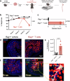
Fig. 2. TF expression is enhanced in the colon and on infiltrating Teffector cells during T cell transfer-induced colitis.
a Schematic diagram of the T cell transfer model of colitis. CD4+ T effector cells were isolated from donor C57BL/6 wild-type (WT) mice and i.p. injected into host Rag1−/− mice (Rag1−/− T cells). Rag1−/− mice i.p. injected with PBS were used as a control (Rag1−/− vehicle). b Colitis developed over a 4-week period, and disease progression was measured by % weight loss compared to original weight. c This was confirmed by subsequent colon histology analysis. d TF staining and e Corrected total fluorescence (CTF) of mice colons following T cell transfer-induced colitis. f TF and CD3 co-staining of mouse colons following T cell transfer-induced colitis (reproduced n = 4 Rag1−/− T cells and n = 3 Rag1−/− vehicle). 2-way ANOVA (b) or two-tailed Student’s t-test (c, e) was used to determine statistical significance from 3–4 biological replicates, n = 4 male Rag1−/− T cells and n = 3 male Rag1−/− vehicle, expressed as mean ± s.e.m. Scale bars: 100 µm (d, f). Source data are provided in the Source Data file. Created in BioRender. Preston, R. (2025) https://BioRender.com/o91u731.
Fig. 3
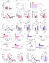
Fig. 3. T cell activation enhances TF-mediated CD4+ T cell thrombogenicity.
CD4+ T cells were isolated from donor human blood and plated at a density of 0.8 × 10⁶/ml with IL-2 for unactivated conditions (θ). Plated cells were activated by anti-CD3/anti-CD28 activation beads and stimulated with IL-2 for Th0 conditions, and differentiation cytokines (αIL-4 + IL-12) were added to skew cells to a Th1 lineage. ad Following 5 days in culture, θ, Th0 and Th1 cells were washed with EDTA-containing PBS, and their ability to initiate thrombin generation was analysed by calibrated automated thrombinography in normal pooled platelet-poor plasma. b Lagtime, c peak thrombin levels and d endogenous thrombin potential (ETP) were measured and compared between θ, Th0 and Th1 cells. e The rate of clot formation was measured in θ, Th0 and Th1 cells. fi θ, Th0 and Th1 cell-mediated thrombin generation was analysed by calibrated automated thrombinography using FVII-deficient platelet-poor plasma. g Lagtime, h peak thrombin levels and i ETP were measured and compared between θ, Th0 and Th1 cells. j F3 gene expression, k, l the percentage of cells expressing TF, m cell surface TF expression and n T cell-dependent FXa generation was measured in θ, Th0 and Th1 cells. Student’s paired t-test (two-tailed) (ce, gi, ln), Wilcoxon test (two-tailed) (b), or Mann–Whitney U Test (two-tailed) (j) was used to determine statistical significance. Data is expressed as mean ± s.d. (be, gi, ln) for 7 (ad), 10 (e), 6 (gi), 8 (j) and 4 (ln) biological donors/group. Source data are provided in the Source Data file.
Fig. 4
Fig. 4. CD4+ T cell activation upregulates markers of TF decryption.
CD4+ T cells were isolated from donor human blood and skewed to θ (unactivated cells), Th0, and Th1 cell subtypes as described previously. a ASMase translocation to the cell surface, d PS exposure on the outer membrane leaflet, and g PDI recruitment to the cell surface were assessed. TF decryption pathways in θ, Th0 and Th1 were analysed by b, c ASMase cell surface expression, e, f fluorescently labelled lactadherin binding to exposed cell surface PS, and h, i cell surface PDI expression. Student’s paired t-test (two-tailed) (c, f, i) was used to determine statistical significance. Data is expressed as mean ± s.d. (c, f, i) for 5 (c), 8 (f), and 4 (i) biological donors/group. Source data are provided in the Source Data file. Created in BioRender. Preston, R. (2025) https://BioRender.com/x11c079.
Fig. 5
Fig. 5. Peripheral CD4+ T cells from IBD patients exhibit TF-mediated thrombogenicity.
CD4+ T cells were isolated from donor adult human peripheral blood, inflamed non-IBD paediatric peripheral blood and paediatric IBD peripheral blood were plated at a density of 1 × 10⁶/ml and incubated for 24–48 h at 37 °C in AIM-V media supplemented with immune replacement serum. ad The percentage of cells expressing TF and e their cell surface TF expression was measured by flow cytometry and compared between groups. fj Plated cells were washed in EDTA-containing PBS, and their ability to initiate thrombin generation was analysed by calibrated automated thrombinography in FXII-deficient plasma. g Lagtime, h peak thrombin levels and i ETP were measured. Furthermore, the ability of isolated T cells to facilitate FXa generation in which the T cells were the sole source of TF, was measured (j). Mann–Whitney U Test (two-tailed) (d, e, g) or Student’s t-test (two-tailed) (j, h, i) was used to determine statistical significance. Data is expressed as mean ± s.e.m. (d, e, gj) for d, e, j 17 biological donors (n = 12 IBD, n = 2 inflamed non-IBD, N = 3 healthy adult), gi 21 biological donors (n = 13 IBD, n = 2 inflamed non-IBD, N = 6 healthy adult) and j 18 biological donors (n = 12 IBD, n = 2 inflamed non-IBD, N = 4 healthy adult). Source data are provided in the Source Data file.
Fig. 6
Fig. 6. Activated PC is downregulated in IBD and limits thrombogenic CD4+ T cell activity.
a PROC (Protein C; PC) expression in colonic biopsies from patients in the RISK study (GEO ID GSE57945) (iCD, n = 162, cCD, n = 56, UC, n = 62, control non-IBD group, n = 42). b, c Following cell culture, T cells were washed and incubated with fluorescently labelled activated PC. Binding was measured by flow cytometry. Following activated PC pre-treatment, activated PC was removed, θ (unactivated cells) and Th0 cells were washed with EDTA-containing PBS, and their capacity to initiate clotting was analysed by dg thrombin generation and h clot formation assays. Activated PC pre-treated Th1 cell-dependent thrombin generation was analysed by il thrombin generation assays and m clot formation assays. Following activated PC pre-treatment, Th0 procoagulant activity was analysed by n FXa generation assay, o F3 gene expression and r, s PDI cell surface expression by flow cytometry. Similarly, Th1 cell thrombogenicity was analysed by p FXa generation assay, q F3 gene expression and t, u PDI cell surface expression by flow cytometry. The contribution of PDI-mediated TF decryption was assessed by treating Th0 (v, w) and Th1 (x, y) cells with 10 mM rutin for 1 h. Cells were washed with EDTA-containing PBS, and their capacity to initiate clotting was analysed by vy thrombin generation. Student’s t-test (two-tailed) (c), Mann–Whitney U Test (two-tailed) (a, nq), Student’s paired t-test (two-tailed) (eg, jl, m, s, u, w, y), or Wilcoxon test (h) was used to determine statistical significance. Data is expressed as mean ± s.e.m. (a, c, eh, jl, mq, s, u, w, y) for a 322 biological donors (iCD, n = 162, cCD, n = 56, UC, n = 62, control non-IBD group, n = 42), c 5–10 biological donors (n = 5 θ, n = 10 Th0 and n = 9 Th1), eg, jl 6 biological donors, h, m 10 biological donors, n, p, s, u 4 biological donors, o, q 8 biological donors and w, y 3–5 biological donors (n = 3 θ, n = 5 Th0/Rutin and n = 5 Th1/Rutin). Source data are provided in the Source Data file.
Fig. 7
Fig. 7. Activated PC inhibits TF-mediated T cell thrombogenicity in IBD patient peripheral CD4+ T cells.
CD4+ T cells were isolated from donor adult human peripheral blood, inflamed non-IBD paediatric peripheral blood and paediatric IBD peripheral blood, plated at a density of 1 × 10⁶/ml and incubated at 37 °C in AIM-V media supplemented with immune replacement serum, +/- 20 nM of activated PC for 24–48 h. ad Cells were washed in EDTA-containing PBS, and their ability to initiate thrombin generation was analysed by calibrated automated thrombinography in FXII-deficient plasma. b Lagtime, c ETP and d peak thrombin levels were measured and compared, as was their ability to facilitate FXa generation (e). Mann–Whitney U Test (two-tailed) (bd) or Student’s t-test (two-tailed) (e) was used to determine statistical significance for all conditions except IBD vs IBD + activated PC. For these conditions, either the Wilcoxon test (two-tailed) (bd) or the Student’s paired t-test (two-tailed) (e) was used as the data is matched. Data is expressed as mean ± s.e.m. for bd 21 biological donors (n = 13 IBD/IBD activated PC, n = 2 inflamed non-IBD, N = 6 healthy adult) and e 19 biological donors (n = 13 IBD, n = 2 inflamed non-IBD, N = 4 healthy adult). Source data are provided in the Source Data file.

References

    1. Lee, S. H., Kwon, J. E. & Cho, M. L. Immunological pathogenesis of inflammatory bowel disease. Intest. Res.16, 26–42 (2018). - PMC - PubMed
    1. European Federation of Crohns & Ulcerative Colitis Associations (EFFCA). What is IBD?https://efcca.org (EFFCA, 2024).
    1. Li, Y. et al. Stem-like T cells are associated with the pathogenesis of ulcerative colitis in humans. Nat. Immunol.25, 1231–1244 (2024). - PMC - PubMed
    1. Alayo, Q. A. et al. Systematic review with meta-analysis: safety and effectiveness of combining biologics and small molecules in inflammatory bowel disease. Crohns Colitis 3604, otac002 (2022). - PMC - PubMed
    1. Kappelman, M. D. et al. Thromboembolic risk among Danish children and adults with inflammatory bowel diseases: a population-based nationwide study. Gut60, 937–943 (2011). - PubMed

MeSH terms

LinkOut - more resources