. 2024 Oct-Dec;65(4):693-712.
doi: 10.47162/RJME.65.4.17.

Biological and cytological-morphological assessment of tuberculous pleural effusions

Affiliations

Biological and cytological-morphological assessment of tuberculous pleural effusions

Iancu Emil Pleşea et al. Rom J Morphol Embryol. 2024 Oct-Dec.

Abstract

Aim: Tuberculosis (TB) came back in the top of causes for infectious disease-related deaths and its pleural involvement is still in the top two extrapulmonary sites. The authors continued their studies on TB pleural effusions (Pl-Effs) with the assessment of biological and cytological variable of pleural fluid (PF), introducing in the investigation algorithm and testing a new tool, the computer-assisted evaluation of cell populations on PF smears.

Patients, materials and methods: A series of 85 patients with TB pleurisy (PLTB) were selected from a larger group of 322 patients with different types of Pl-Effs. The algorithm of investigation included. clinical variables, biological assays of PF, gross aspects including imagistic variables and PF cytology on May-Grünwald-Giemsa (MGG)-stained smears. All the data obtained were entered into and processed using Microsoft Excel module of the 2019 Microsoft Office Professional software along with the 2014 XLSTAT add-in program for MS Excel. The PF cellularity was assessed qualitatively by a cytologist and quantitatively with in-house software. Continuous variables were compared using Pearson's correlation test, while categorical variables were compared using χ² (chi-squared) test.

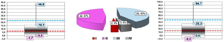
Results: Our analysis showed that patients were usually males, aged between 25 and 44 years with Pl-Eff discovered at clinical imagistic examination, almost always one-sided and free in the pleural cavity. Its extension was either moderate or reduced. The PF had a serous citrine appearance in most of the cases, and biological characteristics pleaded for an exudate [high levels of proteins and lactate dehydrogenase (LDH)], with elevated adenosine deaminase (ADA) values and rich in lymphocytes (Ly). The attempt to identify the pathogen in PF was not of much help. Apart from Ly, neutrophils [polymorphonuclear neutrophils (PMNs)] were a rare presence and their amount had only a trend of direct correlation with Ly. The same situation was encountered in the case of mesothelial cells (MCs). The comparison between the qualitative and the quantitative, computer-assisted evaluations of cytological smears showed that the results of the two methods overlapped in less than one third of the cases, although the sensitivity and specificity values as well as the two calculated predictive values of the qualitative method were encouraging.

Conclusions: The assessment of biological variables and cell populations of the PF are basic tools in the diagnosis of pleural TB. The assessment of PF cell population could be improved by the use of computer-assisted quantitative analysis of the PF smears, which is simple to design, easy to introduce and handle and reliable.

Keywords: biology; cytology; pleura; pleural fluid; tuberculosis.


Conflict of interest statement

The authors declare that they have no conflict of interests.

Figures

Figure 1
Figure 1
Glucose amount: (Left) PF’s glucose values; (Right) Patients’ PF’s glucose distributions. PF: Pleural fluid.
Figure 2
Figure 2
Proteins’ amount: (Left) PF’s proteins values; (Right) Patients’ PF’s proteins distributions. PF: Pleural fluid
Figure 3
Figure 3
LDH’s amount: (Left) PF’s LDH values; (Right) Patients’ PF’s LDH distributions. LDH: Lactate dehydrogenase; PF: Pleural fluid
Figure 4
Figure 4
ADA’s amount: (Left) PF’s ADA values; (Right) Patients’ PF’s ADA distributions. ADA: Adenosine deaminase; PF: Pleural fluid
Figure 5
Figure 5
Results of PF cultures
Figure 6
Figure 6
Sides of PF: (a) Right pleural cavity; (b) Bilateral involvement; (c) Left pleural cavity
Figure 7
Figure 7
Distribution of PF in pleural cavities. B: Bilateral; L: Left; R; Right
Figure 8
Figure 8
Types of PF extension: (a) Minimal extension; (b) Moderate extension; (c) Massive extension; (d) Hemithorax extension; (e) Loculated; (f) Locked up
Figure 9
Figure 9
Assessment of PF extension on Rx
Figure 10
Figure 10
PF’s amount: (Left) PF’s extracted amounts values; (Right) Patients’ PF’s extracted amounts distributions
Figure 11
Figure 11
The amount of PF evacuated: (a) Reduced; (b) Moderate; (c) Massive
Figure 12
Figure 12
Different gross aspects of PF evacuated: (a) Serous–citrine; (b) Hemorrhagic; (c) Serous–hemorrhagic; (d) Purulent.
Figure 13
Figure 13
Distribution of PF gross aspects. H: Hemorrhagic; P: Purulent; Se-Ci: Serous-citrine; Se-H: Serous-hemorrhagic
Figure 14
Figure 14
Qualitative assessment of PF cell population
Figure 15
Figure 15
Qualitative assessment of PF smears: (a) Cellularly very rich; (b) Cellularly poor; (c) Frequent cells; (d) Cellularly rich. MGG staining: (a, c and d) ×40; (b) ×20. MGG: May-Grünwald–Giemsa; PF: Pleural fluid
Figure 16
Figure 16
Distribution of cytological diagnosis types
Figure 17
Figure 17
Cytological diagnoses of PF smears: (a) Normal smear; (b) Chronic inflammation + mesothelial reaction; (c) “Warmed-up” chronic inflammation; (d) Hemorrhagic smear + chronic inflammation. MGG staining: (a–d) ×40
Figure 18
Figure 18
PF smears cellularity: (Left) Cells/×40 field values; (Right) Distribution depending on cell density/×40 field.
Figure 19
Figure 19
Qualitative assessment of PF smears: (a) Normal smear; (b) Cellular smear; (c) Hypercellular smear; (d) Hypocellular smear. MGG staining: (a–d) ×40
Figure 20
Figure 20
PF smears Ly population: (Left) Absolute lymphocytes count; (Center) Distribution depending on Ly percentage from nucleated cells/×40 field; (Right) Percentage of Ly from nucleated cells. I: ≥90%; II: ≥50%; III: <50%. Ly: Lymphocytes; PF: Pleural fluid
Figure 21
Figure 21
Lymphocytes’ density in patients with pathological smears: (a) >90%; (b) <50%. MGG staining: (a and b) ×40.
Figure 22
Figure 22
PMNs in patients with pathological smears: (a) Minimum number of PMNs; (b) Maximum number of PMNs. MGG staining: (a and b) ×40
Figure 23
Figure 23
PF smears PMN population: (Left) Absolute PMNs count/×40 field; (Center) Distribution depending on PMNs percentage from nucleated cells; (Right) Number of PMNs from WBCs/×40 field. I: >50%; II: >20%; III: >2%; IV: >0%; V: 0%
Figure 24
Figure 24
Mesothelial cells density in patients with pathological smears: (a) No MCs on the ×40 field; (b) 47 MCs/×40 field. MGG staining: (a and b) ×40
Figure 25
Figure 25
Mesothelial cells density in patients with pathological smears: (Left) Absolute MCs count/×40 field; (Center) Distribution depending on MCs percentage from nucleated cells; (Right) Number of MCs from nucleated cells/×40 field. I: >80%; II: >5% – <40%; III: >1% – <5%; IV: >0% – <1%
Figure 26
Figure 26
RBCs’ density in patients with pathological smears: (Left) Absolute RBCs count/×40 field; (Center) Distribution depending on RBCs percentage from nucleated cells; (Right) Number of RBCs from all cells/×40 field. I: >50%; II: >10% – <50%; III: <10%
Figure 27
Figure 27
RBCs’ density in patients with pathological smears: (a) No RBCs on the ×40 field; (b) 496 RBCs/×40 field

References

    1. Global Tuberculosis Programme (GTB) . Global Tuberculosis Report 2024 . Geneva, Switzerland : World Health Organization (WHO) ; 2024 .
    1. Kang W, Yu J, Du J, Yang S, Chen H, Liu J, Ma J, Li M, Qin J, Shu W, Zong P, Zhang Y, Dong Y, Yang Z, Mei Z, Deng Q, Wang P, Han W, Wu M, Chen L, Zhao X, Tan L, Li F, Zheng C, Liu H, Li X, A E, Du Y, Liu F, Cui W, Wang Q, Chen X, Han J, Xie Q, Feng Y, Liu W, Tang P, Zhang J, Zheng J, Chen D, Yao X, Ren T, Li Y, Li Y, Wu L, Song Q, Yang M, Zhang J, Liu Y, Guo S, Yan K, Shen X, Lei D, Zhang Y, Yan X, Li L, Tang S. The epidemiology of extrapulmonary tuberculosis in China: a large-scale multi-center observational study. PLoS One. 2020;15(8):e0237753–e0237753. - PMC - PubMed
    1. Kim S, de Los, Jung E. Country-specific intervention strategies for top three TB burden countries using mathematical model. PLoS One. 2020;15(4):e0230964–e0230964. - PMC - PubMed
    1. Vorster MJ, Allwood BW, Diacon AH, Koegelenberg CFN. Tuberculous pleural effusions: advances and controversies. J Thorac Dis. 2015;7(6):981–989. - PMC - PubMed
    1. Golli AL, Niţu MF, Turcu F, Popescu M, Ciobanu-Mitrache L, Olteanu M. Tuberculosis remains a public health problem in Romania. Int J Tuberc Lung Dis. 2019;23(2):226–231. - PubMed

LinkOut - more resources