. 2025 Jan 24;14(1):239-259.
doi: 10.21037/tlcr-2024-1200. Epub 2025 Jan 22.

GNGT1 remodels the tumor microenvironment and promotes immune escape through enhancing tumor stemness and modulating the fibrinogen beta chain-neutrophil extracellular trap signaling axis in lung adenocarcinoma

Affiliations

GNGT1 remodels the tumor microenvironment and promotes immune escape through enhancing tumor stemness and modulating the fibrinogen beta chain-neutrophil extracellular trap signaling axis in lung adenocarcinoma

Lin-Lin Fan et al. Transl Lung Cancer Res. .

Abstract

Background: Despite the recent advancements in the treatment of cancer, the 5-year survival of patients with non-small cell lung cancer (NSCLC) remains unsatisfactory. Lung adenocarcinoma (LUAD) is NSCLC's most common subtype, and metastasis is the major cause of death in patients with cancer. Therefore, identifying novel targets associated with metastasis in NSCLC is crucial to improving treatment. This study aimed to characterize the expression of GNGT1 in LUAD and to clarify the mechanism underlying the association between the higher expression level of GNGT1 and worse prognosis in patients.

Methods: The transcriptome datasets and clinical information of patients with LUAD were obtained from The Cancer Genome Atlas (TCGA) and Gene Expression Omnibus (GEO) database. Bioinformatics analyses were performed in 515 patients who were stratified into two groups (high- and low-GNGT1 expression group) according to the GNGT1 level. Overall survival, DNA promotor methylation, immune cell infiltration, gene set enrichment analysis (GSEA), and Gene Ontology (GO) and Kyoto Encyclopedia of Genes and Genomes (KEGG) pathway enrichment analyses were performed to elucidate the functions of GNGT1 and to identify the related hub genes in LUAD. Their expression and functions in LUAD were verified using tissues from patients and transgenic mice overexpressing GNGT1 under the control of a lung-specific promoter (Scgb1a1-Cre).

Results: GNGT1 was overexpressed in patients with LUAD and was associated with poor prognosis. GNGT1 expression was significantly correlated with gene alteration and hypomethylated promoter status. High GNGT1 expression in patients with LUAD was associated with advanced lymph node metastasis and the degree of immune cell infiltration. Functional enrichment analyses indicated that differentially expressed genes (DEGs) in the high-GNGT1 group participated in DNA replication, DNA replication preinitiation, and M phase, while cell adhesion molecules, apoptosis, and natural killer cell-mediated cytotoxicity were all downregulated. Messenger RNA and protein levels were correspondingly regulated in human LUAD tissues and the Scgb1a1-Cre; LSL-GNGT1 mouse model (GNGT1fl/+ mice).

Conclusions: GNGT1 was associated with tumor cell proliferation via the enhancement of tumor cell stemness and interaction with driver genes. Elevated GNGT1 expression promoted epithelial-mesenchymal transformation, remodeled the tumor microenvironment, and led to tumor metastasis, ultimately worsening the survival-related prognosis of patients with LUAD.

Keywords: GNGT1; neutrophils extracellular traps (NETs); stemness; tumor microenvironment (TME).


Conflict of interest statement

Conflicts of Interest: All authors have completed the ICMJE uniform disclosure form (available at https://tlcr.amegroups.com/article/view/10.21037/tlcr-2024-1200/coif). R.A.d.M. serves as an unpaid editorial board member of Translational Lung Cancer Research from January 2024 to December 2025. The other authors have no conflicts of interest to declare.

Figures

Figure 1
Figure 1
Identification of DEGs and hub genes in LUAD. (A) Volcano plot of the DEGs between NSCLC tissues and normal lung tissues in the GSE7670, GSE81089, and GSE118370 datasets. (B) Venn diagram of DEGs from the three GEO databases. (C) Volcano plot of DEGs between LUAD tissues and normal lung tissues of TCGA. (D) Venn diagram of hub genes in GEO and the top 300 DEGs of TCGA. (E) KEGG and GO functional enrichment analysis of DEGs between LUAD and adjacent tissues. (F) Overall survival diagram of hub genes in LUAD. (G,H) Expression of differential hub genes between LUAD tissues and normal lung tissues from TCGA. ***, P<0.001. FC, fold change; TCGA, The Cancer Genome Atlas; GEO, Gene Expression Omnibus; BP, biological process; MF, molecular function; KEGG, Kyoto Encyclopedia of Genes and Genomes; HR, hazard ratio; CI, confidence interval; TPM, transcripts per kilobase million; DEGs, differentially expressed genes; LUAD, lung adenocarcinoma; NSCLC, non-small cell lung cancer; GO, Gene Ontology.
Figure 2
Figure 2
GNGT1 expression levels and clinical pathological features in LUAD. (A) Expression levels of GNGT1 of various cancers in TCGA database. (B,C) Expression of GNGT1 in LUAD tissues versus adjacent tissues in (B) GSE30219 and (C) GSE10072. (D) Relative expression levels of mRNA in frozen LUAD tissues and adjacent tissues of patients. (E) Clinical pathological features of GNGT1 expression in TCGA. (F) TP53 mutant status of GNGT1 expression in TCGA. (G) Overall survival analysis of GSE41271 according to GNGT1 expression. (H) Expression levels of GNGT1 protein in formalin-fixed tissues of patients with LUAD with paired normal tissues. (I,J) Fisher exact test of the relationship between GNGT1 immunohistochemical expression and the clinicopathological characteristics of patients with LUAD. ns, P>0.05; *, 0.01
Figure 3
Figure 3
The function analysis of GNGT1 in LUAD. (A) Volcano map of transcriptome analysis of DEGs between the GNGT1 high and low group. (B) Analysis of the top 5 genes associated with GNGT1. (C,D) Expression of the top 5 genes associated with GNGT1 in (C) LUAD and (D) LUSC according to GNGT1 expression. (E,F) GO and KEGG enrichment analysis of GNGT1 associated with (E) upregulation and (F) downregulation in biological pathways. (G) GSEA of genes associated with GNGT1 high expression versus those associated genes with low expression. (H) Relevance of GNGT1 across 14 functional states in difference cancers of cancer in the CancerSEA database. (I,J) The correlation between GNGT1 and functional states (metastasis, inflammation, and quiescence) in LUAD. *, 0.01
Figure 4
Figure 4
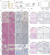
Construction and verification of genetically modified GNGT1fl/+ mice. (A) Pattern plot of conditional overexpression of GNGT1 in lung tissue. (B) Flowchart for obtaining GNGT1fl/+ mice. (C) Expression level of GNGT1 mRNA in multiple organs (lung, liver, kidney, and colon) of GNGT1fl/+ and wild-type mice. (D) Morphologic structure and GNGT1 expression of different organs in GNGT1fl/+ and wild-type mice. (E-G) Expression of cell proliferation markers (E) Ki-67, (F) PCNA, (G) and apoptosis marker caspase-3 in the lungs of GNGT1fl/+ and wild-type mice. ***, P<0.001. ATG, translation initiation codon ATG; Cre, cyclization recombination enzyme; mRNA, messenger RNA; WT, wild type; H&E, hematoxylin and eosin; IHC, immunohistochemistry; PCNA, proliferating cell nuclear antigen.
Figure 5
Figure 5
High expression of GNGT1 promoted stemness gene expression in the lung. (A) The correlation analysis between GNGT1 and the stemness genes of LUAD in TCGA. (B) RT-qPCR and (C) immunofluorescence analysis results of stemness gene expression in lung tissues of wild-type and GNGT1fl/+ mice (×400 magnification). **, 0.001<P<0.05; ***, P<0.001. TPM, transcripts per kilobase million; mRNA, messenger RNA; WT, wild type; DAPI, 4',6-diamidino-2-phenylindole; LUAD, lung adenocarcinoma; TCGA, The Cancer Genome Atlas; RT-qPCR, real-time quantitative polymerase chain reaction.
Figure 6
Figure 6
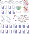
Functional verification of GNGT1 in regulating tumor cell function and reshaping the tumor microenvironment. (A) The correlation analysis between GNGT1 and common driver genes of LUAD in TCGA. (B) Heatmap of the correlation between GNGT1 and driver genes. (C) RT-qPCR validation of driver gene expression level in the wild-type and GNGT1fl/+ mouse models. (D) Venn diagram of GNGT1-related hub genes calculated by cytohubba app in Cytoscape. (E) Heatmap and (F) respective correlation analysis chart between GNGT1 and the obtained key genes. (G) RT-qPCR validation of hub genes level in the wild-type and GNGT1fl/+ mouse models. (H) Model map of FGB-induced NET release and the function of NETs in regulating tumor function and remodeling the microenvironment. (I) qPCR analysis results of the expression of NET-related genes (PADI3, PADI4, and ELANE), three inflammation markers (IL-6, CXCL4, and CXCL15), and the molecules associated with tumor matrix remodeling (HMGB-1 and MMP9). ns, P>0.05; *, 0.01<P<0.05; **, 0.001<P<0.05; ***, P<0.001. TPM, transcripts per kilobase million; mRNA, messenger RNA; WT, wild type; MCC, maximal clique centrality; EPC, edge percolated component; FGB, fibrinogen beta chain; ALB, albumin; CTAG2, cancer/testis antigen 2; AFP, alpha-fetoprotein; HP, haptoglobin; FGF4, fibroblast growth factor 4; CXCL4, platelet factor 4; CXCL15, chemokine (C-X-C motif) ligand 15; HGMB-1, high mobility group protein B1; MMP9, matrix metallopeptidase 9; PAD, peptidyl arginine deiminase; ELANE, elastase; NET, neutrophils extracellular trap; TME, tumor microenvironment; EMT, epithelial-mesenchymal transition; LUAD, lung adenocarcinoma; TCGA, The Cancer Genome Atlas; RT-qPCR, real time quantitative polymerase chain reaction.
Figure 7
Figure 7
Immune infiltration analysis in LUAD and the association with GNGT1 expression. (A) The correlation between GNGT1 gene alteration and the infiltration of different immune cells according to TIMER. (B-D) The correlation between GNGT1 expression and the infiltration of different immune cells as calculated by R software. (E) Immunohistochemical detection of common immune cell markers in LUAD with different GNGT1 expression levels, T-lymphocyte markers CD8 and CD4, neutrophil marker MPO, and B-lymphocyte marker CD20 (red arrow: immune cells; blue arrow: MPO staining of neutrophils was positive). *, 0.01<P<0.05; **, 0.001<P<0.05; ***, P<0.001. LUAD, lung adenocarcinoma; aDC, activated dendritic cell; DC, dendritic cells; iDC, immature dendritic cells; NK, natural killer; pDC, plasmacytoid dendritic cells; TPM, transcripts per kilobase million; H&E, hematoxylin and eosin; MPO, myeloperoxidase; TIMER, Tumor Immune Estimation Resource.

References

    1. Bray F, Laversanne M, Sung H, et al. Global cancer statistics 2022: GLOBOCAN estimates of incidence and mortality worldwide for 36 cancers in 185 countries. CA Cancer J Clin 2024;74:229-63. 10.3322/caac.21834 - DOI - PubMed
    1. Mazzone PJ, Lam L. Evaluating the Patient With a Pulmonary Nodule: A Review. JAMA 2022;327:264-73. 10.1001/jama.2021.24287 - DOI - PubMed
    1. Hu J, Sánchez-Rivera FJ, Wang Z, et al. STING inhibits the reactivation of dormant metastasis in lung adenocarcinoma. Nature 2023;616:806-13. 10.1038/s41586-023-05880-5 - DOI - PMC - PubMed
    1. Spicer JD, Cascone T, Wynes MW, et al. Neoadjuvant and Adjuvant Treatments for Early Stage Resectable NSCLC: Consensus Recommendations From the International Association for the Study of Lung Cancer. J Thorac Oncol 2024;19:1373-414. 10.1016/j.jtho.2024.06.010 - DOI - PubMed
    1. Passaro A, Brahmer J, Antonia S, et al. Managing Resistance to Immune Checkpoint Inhibitors in Lung Cancer: Treatment and Novel Strategies. J Clin Oncol 2022;40:598-610. 10.1200/JCO.21.01845 - DOI - PubMed