. 2025 Feb 11:20:1843-1864.
doi: 10.2147/IJN.S500810. eCollection 2025.

Silica Nanoparticles Loaded With Selenium Quantum Dots Reduce Myocardial Ischemia-Reperfusion Injury by Alleviating Ferroptosis and Mitochondrial Dysfunction

Affiliations

Silica Nanoparticles Loaded With Selenium Quantum Dots Reduce Myocardial Ischemia-Reperfusion Injury by Alleviating Ferroptosis and Mitochondrial Dysfunction

Taixi Li et al. Int J Nanomedicine. .

Abstract

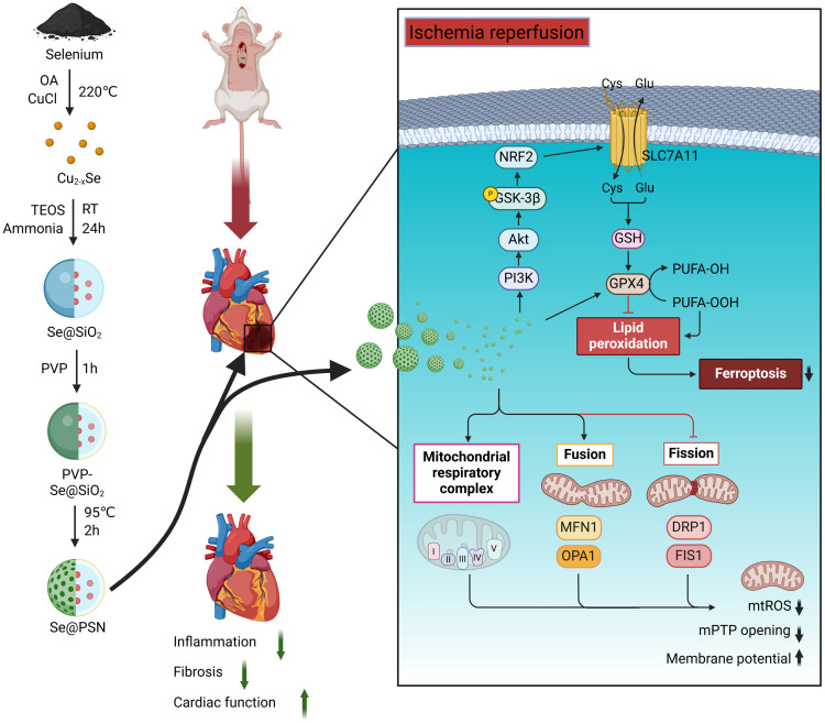
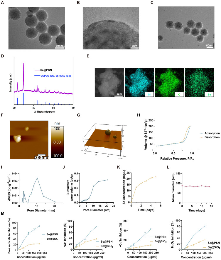
Purpose: Myocardial ischemia-reperfusion (IR) injury, a significant challenge in cardiovascular treatment, is primarily driven by ferroptosis and mitochondrial dysfunction. Despite extensive research, no clinical therapies effectively target ferroptosis in IR injury. This study aims to develop selenium-quantum-dot-loaded porous silica nanospheres (Se@PSN) as a novel therapeutic approach to address IR injury.

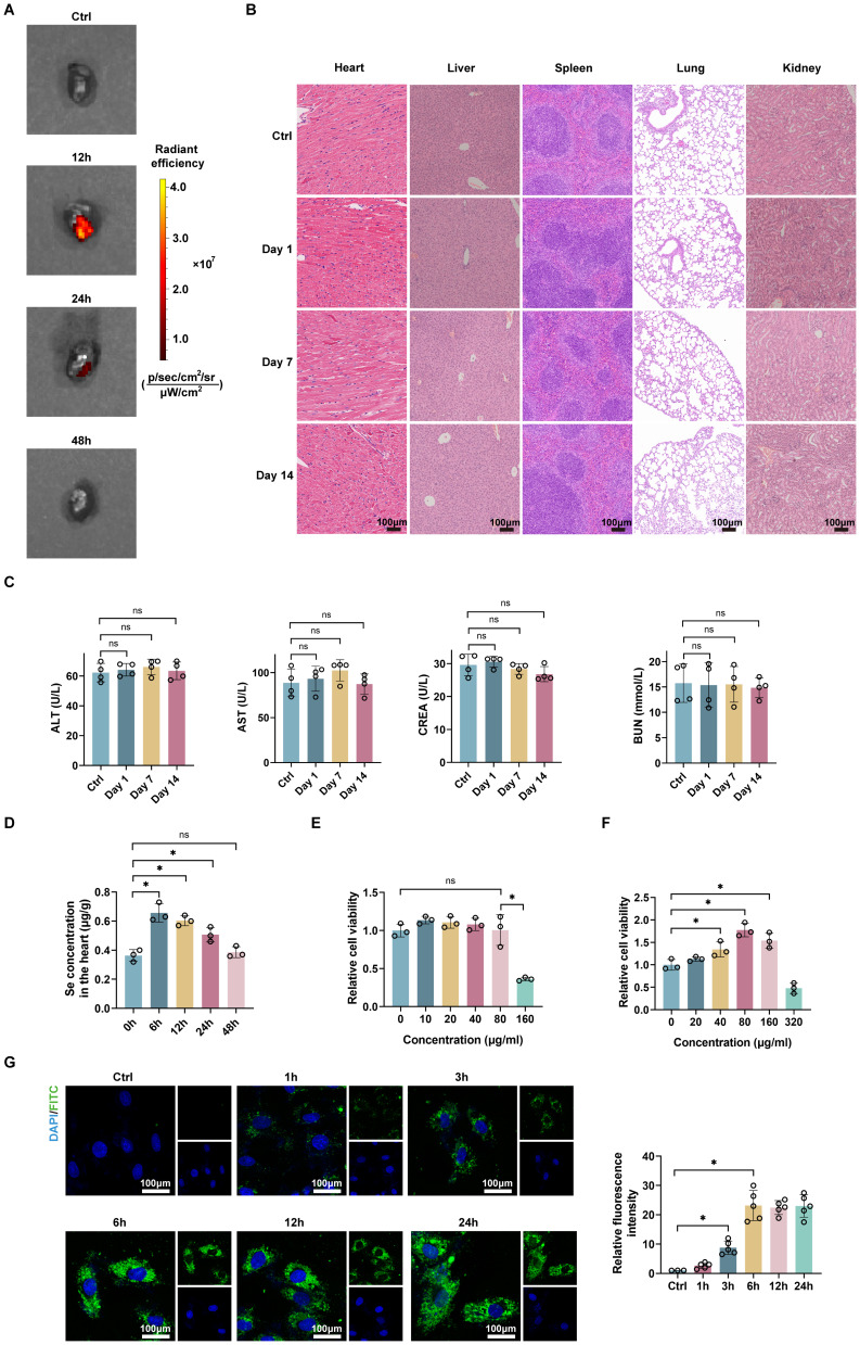
Patients and methods: Se@PSN were synthesized and tested for their reactive oxygen species (ROS) scavenging capabilities and biocompatibility. Additionally, the effects of Se@PSN on ferroptosis, mitochondrial damage, oxidative stress, and myocardial IR injury severity were evaluated.

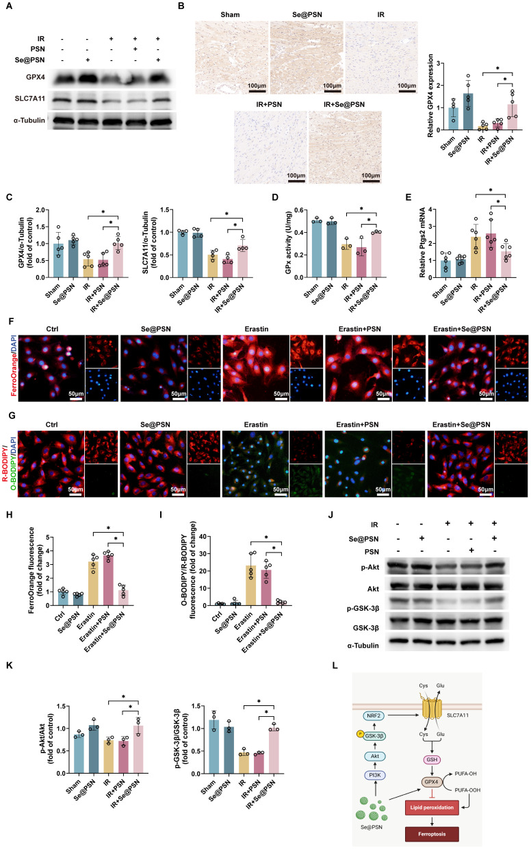
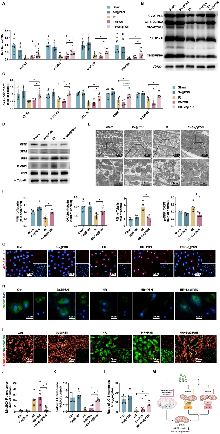
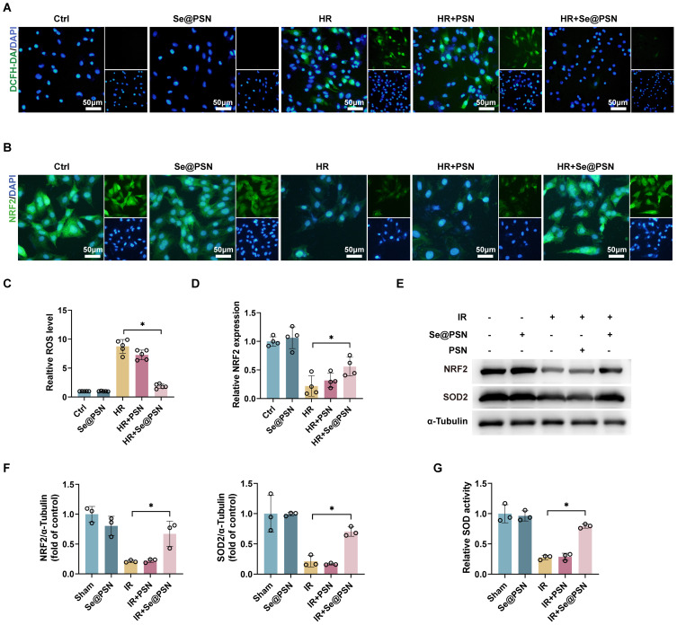
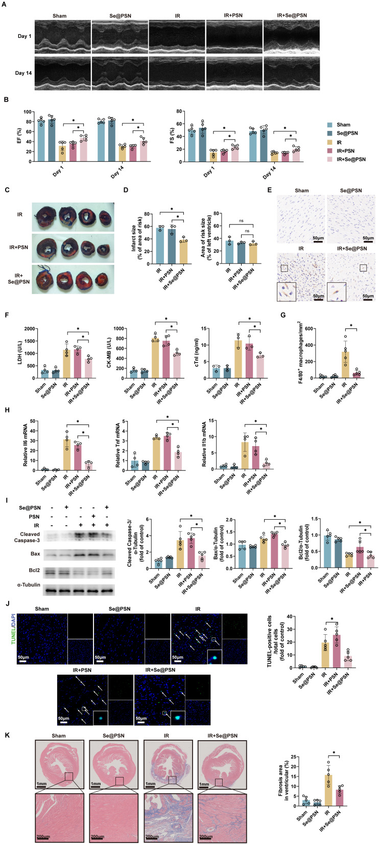
Results: Se@PSN enhanced the stability of selenium quantum dots and exhibited strong ROS scavenging abilities. Additionally, Se@PSN exhibited excellent biocompatibility. The Se@PSN treatment increased GPX4 levels, effectively inhibiting ferroptosis in cardiomyocytes. Furthermore, Se@PSN promoted the expression of mitochondrial respiratory complexes, mitigating oxidative phosphorylation damage and preserving mitochondrial function. These effects collectively resulted in reduced myocardial loss, inflammation, and fibrosis following IR injury. Compared to PSN alone, Se@PSN showed superior therapeutic efficacy against IR injury.

Conclusion: Se@PSN exhibit great potential in reducing ferroptosis and protecting mitochondrial function, making them a promising therapeutic approach for the treatment of myocardial IR injury.

Keywords: ferroptosis; myocardial ischemia reperfusion; porous silica nanospheres; reactive oxygen species; selenium.


Conflict of interest statement

The authors report no conflicts of interest in this work.

Figures

None
Graphical abstract
Figure 1
Figure 1
Characterization of Se@PSN. (A and B) Low and high magnification TEM images of solid Se@SiO2. (C) TEM image of Se@PSN. (D) XRD patterns of Se@PSN and standard selenium phase. (E) Elemental mapping showing the distribution of Si, O, and Se. (F and G) AFM analysis of Se@PSN, with height and 3D surface images. (H) Nitrogen adsorption-desorption isotherms of Se@PSN. I, (J) Pore size distribution and cumulative pore volume of Se@PSN. (K) Release profile of selenium concentration from 8g L−1 Se@PSN. (L) Average hydration diameter of Se@PSN in PBS within 14 days. (M) Free radical and ROS inhibition assays demonstrating the dose-dependent neutralization of total free radicals, •OH, •O2, and H2O2 by Se@PSN and Se@SiO2 (n=3).
Figure 2
Figure 2
Pharmacokinetics and Biocompatibility of Se@PSN. (A) Ex vivo fluorescence imaging of heart treated with IR following Se@PSN administration at different time. (B) H&E staining of heart, liver, spleen, lung, and kidney tissues from control and Se@PSN-treated groups at different time. (C) Serum levels of ALT, AST, CREA, and BUN in control and Se@PSN-treated mice at different time (n=4; ns, p>0.05). (D) Time-dependent selenium concentration in the heart from mice treated with IR following Se@PSN administration (n=3, *p < 0.05). (E) Relative cell viability of cells treated with Se@PSN, as measured by CCK8 assay (n=3, *p < 0.05). (F) Relative cell viability of HR-treated H9c2 cells after treatment with different concentrations of Se@PSN (n=3, *p < 0.05). (G) Fluorescence imaging of FITC-Se@PSN in H9c2 cells at different time points post-treatment (n=3-5, *p < 0.05).
Figure 3
Figure 3
Transcriptomic Analysis of Se@PSN Treatment in IR Injury. (A) Schematic illustration of animal intervention. (B) PCA of gene expression profiles between the IR and IR+Se@PSN groups. (C) Volcano plot of DEGs in the IR+Se@PSN group compared to the IR group. (D) Heatmap showing the expression levels of ferroptosis-related genes in both groups. (E) Heatmap of DEGs associated with mitochondrial inner membrane, OXPHOS, oxidoreductase activity, and response to oxidative stress pathways in both groups. (F) GSEA plot for OXPHOS (IR+Se@PSN group vs IR group). (G) GO analysis of DEGs in the IR+Se@PSN group, highlighting enrichment in BP and CC. (H) KEGG pathway analysis of DEGs in the IR+Se@PSN group, showing significant enrichment in key pathways. (I) PPI network of enriched pathways in the IR+Se@PSN group demonstrating the connection between ferroptosis and other key gene sets.
Figure 4
Figure 4
Effects of Se@PSN on Ferroptosis in IR Injury. A, (C) Western blot images and quantification showing the expression levels of GPX4 and SLC7A11 in sham, Se@PSN, IR, IR+ PSN, and IR+Se@PSN groups (n=4-5, *p < 0.05). (B) Immunohistochemistry staining images and quantification of GPX4 in myocardium from different groups (n=4-5, *p < 0.05). (D) Measurement of GPx activities in myocardium from different groups (n=3, *p < 0.05). (E) Relative Ptgs2 mRNA levels in myocardium from different groups (n=6, *p < 0.05). (F and H) FerroOrange staining showing ferrous ions levels in H9c2 cells from control, Se@PSN, erastin, erastin+PSN, and erastin+Se@PSN groups (n=5, *p < 0.05). (G and I) BODIPY-C11 staining demonstrating lipid peroxidation levels in H9c2 cells from different groups (n=5, *p < 0.05). J, (K) Western blot analysis of phosphorylated and total Akt and GSK-3β in myocardium from different groups (n=3, *p < 0.05). (L) Mechanism by which Se@PSN alleviates ferroptosis. Created in BioRender. Li, T. (2025) https://BioRender.com/y58m263.
Figure 5
Figure 5
Impact of Se@PSN on Mitochondria in IR Injury. (A) Relative mRNA levels of mt-Co1, mt-Nd1, mt-Cytb, and mt-Atp6 in myocardium from sham, Se@PSN, IR, IR+PSN, and IR+Se@PSN groups (n=6, *p < 0.05). B, (C) Western blot images and quantification of representative OXPHOS components (ATP5A, UQCRC2, MTCO1, SDHB, NDUFB8) in myocardium from different groups (n=4, *p < 0.05). D, (F) Western blot images and quantification of mitochondrial dynamics proteins (MFN1, OPA1, FIS1, p-DRP1) in myocardium from different groups (n=5, *p < 0.05). (E) TEM images of mitochondrial structures and myocardial fiber structure in myocardium from different groups. (G and J) MitoSOX staining images and quantitative analysis, demonstrating mitochondrial ROS level changes in H9c2 cells from control, Se@PSN, HR, HR+PSN, and HR+Se@PSN groups (n=5, *p < 0.05). (H and K) Calcein AM staining for mPTP opening in H9c2 cells from different groups, with quantitative analysis of fluorescence intensity (n=5, *p < 0.05). (I and L) JC-1 staining images and quantitative analysis, showing mitochondrial membrane potential changes in H9c2 cells from different groups (n=5, *p < 0.05). (M) Mechanism by which Se@PSN protects mitochondria. Created in BioRender. Li, T. (2025) https://BioRender.com/a41f268.
Figure 6
Figure 6
Antioxidant Activity of Se@PSN in Cardiomyocytes. (A and C) ROS levels in H9c2 cells of the control, Se@PSN, HR, HR+PSN, and HR+Se@PSN groups (n=5, *p < 0.05). (B and D) NRF2 expression in H9c2 cells of different groups (n=4-6, *p < 0.05). (E and F) Western blot analysis of NRF2 and SOD2 expression in the myocardium from Sham, Se@PSN, IR, IR+PSN, and IR+Se@PSN groups (n=3, *p < 0.05). (G) Measurement of SOD activity in the myocardium from different groups (n=3, *p < 0.05).
Figure 7
Figure 7
Cardioprotective Effects of Se@PSN in IR Injury. (A) Echocardiography images of mice from Sham, Se@PSN, IR, IR+PSN, and IR+Se@PSN groups at day 1 and 14 post-IR. (B) EF and FS of hearts at day 1 and 14 post-IR from different groups (n=5, *p < 0.05). (C) TTC & Evans Blue stained heart sections from IR, IR+PSN, and IR+Se@PSN groups. (D) Measurement of infarct and risk area sizes of myocardium from different groups (n=3, *p < 0.05). (E and G) Immunohistochemical staining images and quantification for F4/80 positive cells in myocardium from different groups (n=5, *p < 0.05). (F) Serum levels of cardiac injury markers in mice from different groups (n=3-4, *p < 0.05). (H) Relative mRNA levels of inflammatory cytokine including Il6, Tnfa, and Il1b in myocardium from different groups (n=4, *p < 0.05). (I) Western blot images and quantification of cleaved Caspase-3, Bax, and Bcl2 in myocardial from different groups (n=5, *p < 0.05). (J) TUNEL staining image and quantification of myocardial from different groups (n=5, *p < 0.05). (K) Masson’s trichrome staining of myocardial from different groups and fibrosis area quantification in the left ventricle (n=5, *p < 0.05).

Similar articles

References

    1. Virani SS, Alonso A, Aparicio HJ, et al. Heart disease and stroke statistics-2021 update: a report from the American heart association. Circulation. 2021;143(8):e254–e743. doi:10.1161/CIR.0000000000000950 - DOI - PubMed
    1. Khan MA, Hashim MJ, Mustafa H, et al. Global epidemiology of ischemic heart disease: results from the global burden of disease study. Cureus. 2020;12(7):e9349. doi:10.7759/cureus.9349 - DOI - PMC - PubMed
    1. Tejedor S, Dolz-Pérez I, Decker CG, et al. Polymer conjugation of docosahexaenoic acid potentiates cardioprotective therapy in preclinical models of myocardial ischemia/reperfusion injury. Adv. Healthcare Mater. 2021;10(9):2002121. doi:10.1002/adhm.202002121 - DOI - PubMed
    1. Kajstura J, Cheng W, Reiss K, et al. Apoptotic and necrotic myocyte cell deaths are independent contributing variables of infarct size in rats. Lab Invest. 1996;74(1):86–107. - PubMed
    1. Reed GW, Rossi JE, Cannon CP. Acute myocardial infarction. Lancet. 2017;389(10065):197–210. doi:10.1016/S0140-6736(16)30677-8 - DOI - PubMed

MeSH terms

LinkOut - more resources