. 2024 Aug 30;10(1):43-61.
doi: 10.1016/j.jacbts.2024.08.003. eCollection 2025 Jan.

Empagliflozin in Acute Myocardial Infarction Reduces No-Reflow and Preserves Cardiac Function by Preventing Endothelial Damage

Affiliations

Empagliflozin in Acute Myocardial Infarction Reduces No-Reflow and Preserves Cardiac Function by Preventing Endothelial Damage

Panagiota Efstathia Nikolaou et al. JACC Basic Transl Sci. .

Abstract

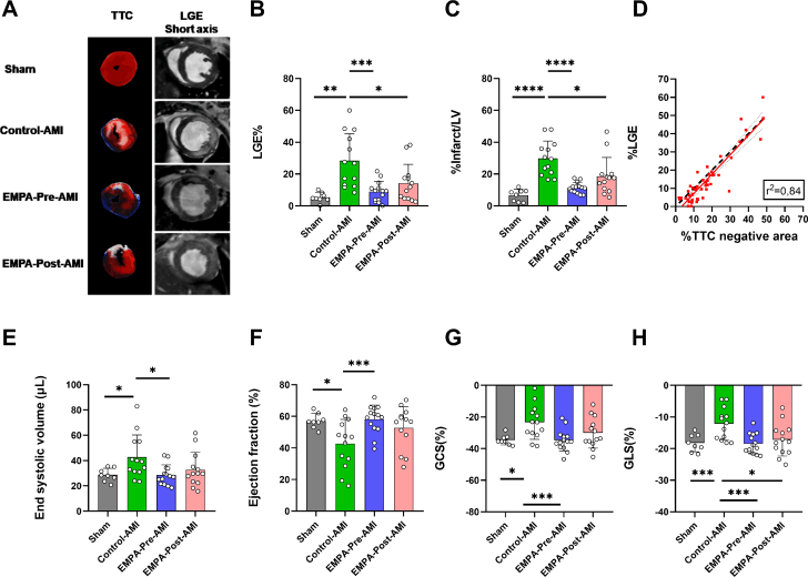
Empagliflozin treatment before acute myocardial infarction mainly targets the endothelial cell transcriptome. Empagliflozin treatment before and after myocardial infarction decreased no reflow and microvascular injury, leading to reduced infiltration of inflammatory cells, reduced infarct size, and improved cardiac function in mice. In diabetic patients receiving empagliflozin after myocardial infarction, perfused boundary region, flow-mediated dilation, and global longitudinal strain were improved.

Keywords: acute myocardial infarction; cardiac magnetic resonance; empagliflozin; microvascular injury; no-reflow.


Conflict of interest statement

The authors acknowledge support of this work by the project “The Greek Research Infrastructure for Personalised Medicine (pMedGR)” (MIS 5002802) under the Action “Reinforcement of the Research and Innovation Infrastructure,” funded by the Operational Programme “Competitiveness, Entrepreneurship and Innovation” (NSRF 2014-2020) and co-financed by Greece and the European Union (European Regional Development Fund). This study was partially supported by an investigator-initiated study from Boehringer Ingelheim to Dr Andreadou. Dr Pieper is an employee of Boehringer Ingelheim Pharma & Co; and provided the research grant to the research institute of Dr Andreadou related to this work. Dr Zuurbier has received a research grant from Boehringer Ingelheim. Dr Nijveldt has received research grants from Philips Volcano and Biotronik; and speaker fees from BMS, Pfizer, and Sanofi Genzyme. Dr van Royen has received research grants from Abbott, Philips, Medtronic, and Biotronik; and speaker fees from Abbott, Bayer, RainMed, and Microport. All other authors have reported that they have no relationships relevant to the contents of this paper to disclose.

Figures

None
Graphical abstract
Figure 1
Figure 1
Effect of AMI and EMPA on Cardiomyocytes, Fibroblasts, and EC Transcriptome 2 Hours After Reperfusion (A) Principal component (PC) analysis plot with the relative gene expression for all samples indicating the discrimination of the cell populations. Heatmap gene expression analyses depicting the significant differences between the Sham and the Control-AMI groups in (B) isolated cardiomyocytes, (C) sorted fibroblasts, and (D) sorted ECs. PC analyses showing the discrimination among the Sham, Control-AMI, and EMPA–Pre-AMI groups in (E) cardiomyocytes, (G) sorted fibroblasts, and (I) sorted ECs. Volcano plots indicating the significant genes as blue dots and the significant and deregulated genes as red dots between the Control-AMI and EMPA–Pre-AMI groups in (F) cardiomyocytes (FDR: <0.10), (H) sorted fibroblasts (FDR: <0.10), and (J) sorted ECs (FDR: <0.05). AMI = acute myocardial infarction; EC = endothelial cells; EMPA = empagliflozin; FDR = false discovery rate.
Figure 2
Figure 2
EMPA Pretreatment Reverses the Deregulated Transcriptome by AMI in ECs (A) Bar plots indicating the significant Reactome pathways between the Control-AMI and EMPA–Pre-AMI group 2 hours after reperfusion in sorted ECs with FDR <0.05. (B) Heatmap analysis demonstrating the 150 genes that are significantly deregulated by AMI and restored by EMPA pretreatment. (C) Experimental protocol for RNA sequencing data validation. Relative gene expression 2 hours after reperfusion as 2−(Cq-CGapdh) (fold change of Control-AMI) for (D) growth factor genes and regulators, (E) genes related to cell adhesion, and (F) genes related to extracellular matrix and collagen degradation. Data are presented as mean ± SD (n = 5-6 per group; 1-way analysis of variance with Tukey post hoc test: ∗P < 0.05; ∗P < 0.05; ∗∗P < 0.01. DMSO = dimethylsulfoxide; FDR = false discovery rate; other abbreviations as in Figure 1.
Figure 3
Figure 3
EMPA Treatment Before and After AMI Induction Reduces No-Reflow and Infarct Size (A) Schematic representation of the experimental protocol. (B) Representative images of Evans Blue and Thioflavin S staining 48 hours after reperfusion. Bar plots for (C) percentage Thioflavin S (%ThioS)–negative area to area at risk, (D) %ThioS–negative area to left ventricle (LV), (E) percentage infarct to area at risk, (F) percentage area at risk to LV, and (G) %ThioS–negative area to infarct size. Data are presented as mean ± SD (n = 8-14 per group; 1-way analysis of variance with Tukey post hoc test: ∗∗P < 0.01; ∗∗∗P < 0.001; ∗∗∗∗P < 0.0001. DMSO = dimethylsulfoxide; NS = normal saline solution; other abbreviations as in Figure 1.
Figure 4
Figure 4
EMPA Treatment Before and After AMI Improves Capillary Integrity and Prevents Infiltration of Inflammatory Cells in the Infarcted Heart (A) Representative images of hematoxylin-eosin staining. Squares indicate the area of focus. Red arrow denotes destroyed/disrupted vessel, and green arrows denote vessels with structural integrity. (B) Representative electron micrographs indicating intramyocardial hemorrhage. Red arrows depict extravasation of erythrocytes, indicating intramyocardial hemorrhage and capillaries clogged by erythrocytes. Yellow arrows denote intact capillaries. (C) Quantification of erythrocytes in transition electron microscopy images and microvascular injury score in hematoxylin-eosin images for the respective groups. (D) Representative images of flow cytometric analysis of the inflammatory cells in the infarcted heart. (E) Bar plots with the percentage of each cell population expressed as a percentage of the nucleated cells. Data are presented as mean ± SD (n = 6-9 per group; 1-way analysis of variance with Tukey post hoc test: ∗P < 0.05; ∗∗P < 0.01; ∗∗∗P < 0.001; ∗∗∗∗P < 0.0001. Abbreviations as in Figure 1.
Figure 5
Figure 5
EMPA Treatment Before and After AMI Reduces Infarct Size and Improves Cardiac Function (A) Representative images of 2,3,5-triphenyl tetrazolium chloride (TTC) staining and late gadolinium enhancement (LGE) in short axis for each experimental group 48 hours after reperfusion. (B) Percentage LGE infarction determined by cardiac magnetic resonance imaging and (C) infarct/left ventricle (LV) via staining. (D) Linear regression (solid red line) and the line of identity (dashed line) comparing the infarct size values determined via staining and %LGE. Bar plots indicating (E) end-systolic volume, (F) LV ejection fraction, (G) global circumferential strain (GCS), and (H) global LV longitudinal strain (GLS). Data are presented as mean ± SD (n = 8-14 per group; 1-way analysis of variance with Tukey post hoc test: ∗P < 0.05; ∗∗P < 0.01; ∗∗∗P < 0.001; ∗∗∗∗P < 0.0001. Abbreviations as in Figure 1.
Figure 6
Figure 6
EMPA Reduces ICAM-1 and MMP-2 Expression and STAT-3 Phosphorylation at Late Reperfusion Representative Western blots (A) 2 hours and (B) 48 hours after reperfusion. Relative densitometric graphs after normalization to Sham of (C) p-(Y703)STAT-3/t-STAT-3, t-STAT-3/β-tubulin, FGF-2/GAPDH, and VEGF/GAPDH 2 hours after reperfusion and (D) p-(Y703)STAT-3/t-STAT-3, t-STAT-3/β-tubulin, FGF-2/GAPDH, and VEGF/GAPDH 48 hours after reperfusion. Representative Western blots (E) 2 hours and (F) 48 hours after reperfusion. Relative densitometric graphs after normalization to Sham of (G) ICAM-1/α-actinin, MMP-2/α-actinin, TIMP-1/β-tubulin, and IGF-1/β-tubulin at 2 hours of reperfusion and (H) ICAM-1/GAPDH, MMP-2/GAPDH, TIMP-1/β-tubulin, and IGF-1/β-tubulin at 48 hours of reperfusion. Dots represent biological replicates. Results are presented as mean ± SD (n = 6 per group; 1-way analysis of variance with Tukey post hoc test: ∗P < 0.05; ∗∗P < 0.01. AMI = acute myocardial infarction; EMPA = empagliflozin; FGF = fibroblast growth factor 2; ICAM = intercellular adhesion molecule; IGF = insulin-like growth factor; MMP = matrix metalloproteinase; STAT = signal transducer and activator of transcription; TIMP = tissue inhibitor of the metalloproteinase; VEGF = vascular endothelial growth factor.
Figure 7
Figure 7
EMPA Treatment Within 2 Months of STEMI Improves Endothelial and Cardiac Dysfunction Altering the Circulating Markers Thrombomodulin and ICAM-1 (A) Schematic representation of the clinical protocol. EMPA was given within 2 months after STEMI, and clinical parameters were evaluated at 4 months and 12 months after inclusion. Box whisker plots with 5th to 95th percentiles indicating (B) perfused boundary region (PBR), (C) pulse-wave velocity (PWV), (D) percentage flow-mediated dilation (FMD), (E) percentage global longitudinal strain (GLS), (F) white blood cell count (WBC), (G) thrombomodulin levels in plasma (ng/mL), and (H) ICAM-1 levels in plasma (mg/mL) at 4- and 12-month follow-ups. Data are presented as mean ± SD (n = 24 for EMPA and n = 18 for Control; 2-way repeated-measures analysis of variance with Tukey post hoc test: ∗P < 0.05; ∗∗∗P < 0.001. % GLS = % global longitudinal strain; EMPA = empagliflozin; FMD% = flow-mediated dilation of the brachial artery; ICAM = intercellular adhesion molecule.

References

    1. Heusch G. Myocardial ischemia/reperfusion: translational pathophysiology of ischemic heart disease. Med. 2024;5:10–31. - PubMed
    1. Heusch G., Gersh B.J. The pathophysiology of acute myocardial infarction and strategies of protection beyond reperfusion: a continual challenge. Eur Heart J. 2017;38:774–784. - PubMed
    1. Hausenloy D.J., Chilian W., Crea F., et al. The coronary circulation in acute myocardial ischaemia/reperfusion injury: a target for cardioprotection. Cardiovasc Res. 2019;115:1143–1155. - PMC - PubMed
    1. Heusch G. The coronary circulation as a target of cardioprotection. Circ Res. 2016;118:1643–1658. - PubMed
    1. Robbers L.F.H.J., Eerenberg E.S., Teunissen P.F.A., et al. Magnetic resonance imaging-defined areas of microvascular obstruction after acute myocardial infarction represent microvascular destruction and haemorrhage. Eur Heart J. 2013;34:2346–2353. - PubMed

LinkOut - more resources