

Hepatic Kaposi Sarcoma after Kidney Transplantation: A Case Report
- PMID: 39958498
- PMCID: PMC11822272
- DOI: 10.3348/jksr.2024.0027
Hepatic Kaposi Sarcoma after Kidney Transplantation: A Case Report
Abstract
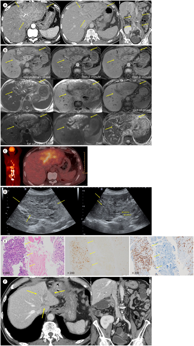
Kaposi sarcoma is an undisputed malignancy associated with a heightened relative risk after transplantation. Similar to other causes of Kaposi's sarcoma, cutaneous involvement is typical in post-transplant patients; however, visceral involvement rarely occurs. We report a rare case of de novo hepatic Kaposi's sarcoma manifesting as an ill-defined infiltrative lesion in the left lobe of the liver in a patient who was immunosuppressed for 9 months after a kidney transplantation using ultrasonography, CT, MRI, and fluorodeoxyglucose-PET.
카포시 육종은 장기이식 후 발병의 상대위험도가 명백히 증가하는 악성 종양 중 하나이다. 다른 원인의 카포시 육종처럼 이식 후 환자에서 발생하는 카포시 육종도 피부 병변의 형태가 가장 흔하지만, 드물게 내장 기관 침범이 발생할 수 있다. 본 증례는 신장 이식 후 9개월간 면역 억제 치료를 받은 환자에서 발견된 간좌엽의 침윤성 병변이 카포시 육종으로 진단된 경우로서, 저자들은 신장 이식 환자에서 드물게 발생하는 간의 카포시 육종 사례를 초음파, CT, MRI 및 fluorodeoxyglucose-PET 영상 소견과 함께 보고하고자 한다.
Keywords: Kaposi Sarcoma; Kidney Transplantation; Liver.
Copyrights © 2025 The Korean Society of Radiology.
Conflict of interest statement
Conflicts of Interest: The authors have no potential conflicts of interest to disclose.
Figures
Similar articles
-
Kaposi's sarcoma in organ transplant recipients. The Collaborative Transplantation Research Group of Ile de France.Eur J Med. 1993 Jun-Jul;2(6):339-43. Eur J Med. 1993. PMID: 8252179
-
Molecularly targeted therapy for Kaposi's sarcoma in a kidney transplant patient: case report, "what worked and what did not".BMC Nephrol. 2007 Mar 27;8:6. doi: 10.1186/1471-2369-8-6. BMC Nephrol. 2007. PMID: 17386117 Free PMC article.
-
[Kaposi's syndrome following transplantation].J Mal Vasc. 1991;16(2):163-5. J Mal Vasc. 1991. PMID: 1861111 French.
-
Vertebral Lesions from AIDS-Related Kaposi's Sarcoma.Curr HIV Res. 2011 Jun;9(4):270-5. doi: 10.2174/157016211796320342. Curr HIV Res. 2011. PMID: 21631426 Review.
-
The diagnostic challenge and management of pulmonary Kaposi's sarcoma in renal transplant recipients.Saudi Med J. 2001 Dec;22(12):1061-4. Saudi Med J. 2001. PMID: 11802176 Review.
References
-
- Organ Procurement and Transplantation Network. 2022 organ transplants again set annual records; organ donation from deceased donors continues 12-year record-setting trend. 2023. [Accessed January 27, 2024]. Available at. https://optn.transplant.hrsa.gov/news/2022-organ-transplants-again-set-a... .
-
- Restrepo CS, Martínez S, Lemos JA, Carrillo JA, Lemos DF, Ojeda P, et al. Imaging manifestations of Kaposisarcoma. Radiographics. 2006;26:1169–1185. - PubMed
Publication types
LinkOut - more resources
Full Text Sources
