Case Reports
. 2025 Jan;86(1):148-153.
doi: 10.3348/jksr.2024.0091. Epub 2025 Jan 20.

Isolated Perihepatic Peritoneal Leiomyoma: A Case Report

Case Reports

Isolated Perihepatic Peritoneal Leiomyoma: A Case Report

Seul Ki Kim et al. J Korean Soc Radiol. 2025 Jan.

Abstract

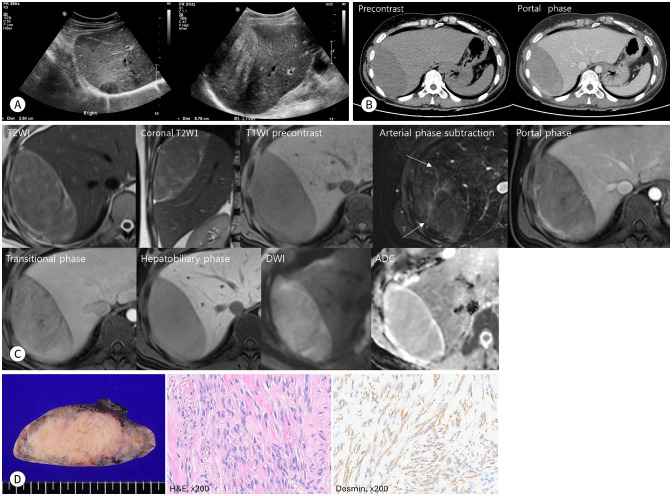
Peritoneal leiomyomas are extremely rare. Most reported cases are that of disseminated peritoneal leiomyomatosis, making isolated leiomyoma an uncommon occurrence. Given that isolated leiomyoma is rare, the preoperative diagnosis of isolated leiomyoma is challenging. To date, very few reports have described the radiological findings of isolated peritoneal leiomyoma. This study aimed to present a rare case of isolated peritoneal leiomyoma in the perihepatic region of a 54-year-old female, and present the US, CT, and MRI based radiological findings of the case.

복막 평활근종은 매우 드물다. 대부분 보고된 사례들은 파종성 복막 평활근종으로, 단일 평활근종은 흔하지 않다. 단일 평활근종은 드물기 때문에 단일 평활근종의 수술 전 진단은 어렵다. 현재까지 단일 복막 평활근종의 영상의학적 소견에 대한 보고는 거의 없다. 본 연구는 54세 여성의 간 주위에서 발견된 드문 단일 복강내 평활근종 증례를 제시하고, 초음파, 컴퓨터단층촬영, 자기공명영상을 기반으로 한 영상학적 소견을 보여주고자 한다.

Keywords: Leiomyoma; Liver; Magnetic Resonance Imaging; Peritoneum; Tomography, X-Ray Computed.


Conflict of interest statement

Conflicts of Interest: The authors have no potential conflicts of interest to disclose.

Figures

Fig. 1
Fig. 1. A 54-year-old female with peritoneal leiomyoma presented with a perihepatic mass.
A. Initial US (left) shows a well-defined hypoechoic mass of approximately 3.5 cm. After 7 years (right), this mass significantly increased to approximately 10 cm and changed to a heterogeneous hyperechoic form. B. Precontrast CT shows a well-defined smooth mass with low attenuation compared to the surrounding liver tissue. Postcontrast CT shows the large, lentiform-appearing perihepatic tumor has a slightly lower degree of contrast enhancement than the adjacent liver parenchyma. C. On MRI, the tumor shows heterogeneously high SI on T2WI and relatively homogenous low SI on pre-T1WI compared to the adjacent liver parenchyma, and early heterogenous and slow progressive enhancement without definite intratumoral necrosis. On DWI with a b-value of 800 s/mm2, the mass shows a subtly high SI and a subtly high ADC value on the ADC map compared to liver parenchyma. The tumor exhibits several prominent intratumoral arterial vascular structures arising from the diaphragmatic side in arterial phase subtraction image (arrows) and a negative beak sign between the tumor and the liver on coronal T2WI indicating an extrahepatic mass. D. The gross surgical specimens appear as well-defined mass with a whitish cut surface (left). Microscopy shows proliferation of spindle cells with elongated nuclei and eosinophilic cytoplasm corresponding to smooth muscle fibers (middle, H&E stain, ×200). Immunohistochemical stain for desmin (right, ×200) is positive. ADC = apparent diffusion coefficient, DWI = diffusion-weighted imaging, H&E = hematoxylin & eosin stain, SI = signal intensity, WI = weighted image

References

    1. Woo J, Choi SY, Kim HK, Lee JE, Lee MH, Lim S. Extremely rare CT and MRI findings of peritoneal leiomyoma mimicking hepatic mass: a case report. J Korean Soc Radiol. 2023;84:946–951. - PMC - PubMed
    1. Micha G, Galatis DG, Kalopita K, Strongylos A, Benekos C, Kalaitzi K, et al. Unusual case of a parasitic extra-uterine leiomyoma presenting with lower abdominal pain. Cureus. 2022;14:e30141. - PMC - PubMed
    1. Fasih N, Prasad Shanbhogue AK, Macdonald DB, Fraser-Hill MA, Papadatos D, Kielar AZ, et al. Leiomyomas beyond the uterus: unusual locations, rare manifestations. Radiographics. 2008;28:1931–1948. - PubMed
    1. Bhargava P, Haque K, Vea R, Turbat-Herrera E, Chu Q, Sangster G, et al. Parasitic leiomyoma presenting as an inguinal hernia in a postmenopausal woman. Radiol Case Rep. 2018;13:767–771. - PMC - PubMed
    1. Yang L, Liu N, Liu Y. Leiomyomatosis peritonealis disseminata: three case reports. Medicine (Baltimore) 2020;99:e22633. - PMC - PubMed

Publication types

LinkOut - more resources