Cranio-cervical hyperpneumatization: a case report
- PMID: 39958674
- PMCID: PMC11829802
- DOI: 10.1093/bjrcr/uaaf003
Cranio-cervical hyperpneumatization: a case report
Abstract
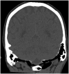
Hyperpneumatization is a rare pathological process where air-filled cavitation form within solid bone architecture occurring at sites where physiological pneumatization is not seen. Extension of this process into the atlanto-occipital region is considered extremely rare and is only quoted several times in the literature. In this case report, we present a 66-year-old man who presented with an 8-month history of a worsening frontal headache and blocked sensation in his left ear. Subsequent CT head evaluation revealed hyperpneumatization affecting C1 vertebra, temporal and occipital bones with extension into the clivus. A rare complication of epidural emphysema was seen. The aetiology of hyperpneumatization is uncertain, although it is thought to be either congenital or acquired. In our case, clinical suggestion of eustachian tube dysfunction and radiological findings of thickened sinus mucosa and a unilateral nasal polyp point to chronic recurrent coryzal illnesses, which may indicate an acquired mechanism. Management is mostly conservative with surgical management reserved for high risk or refractory cases.
Keywords: Hyperpneumatisation; aquired; atlanto-occipital; epidural; neuroradiology; pneumorrhacis; valsalva.
© The Author(s) 2025. Published by Oxford University Press on behalf of the British Institute of Radiology.
Conflict of interest statement
None declared.
Figures
References
-
- AbdulAzeez MM, Huber P-Z, Alsaadi SB, Vladimir C-NB, Salazar LRM, Hoz SS. Cranio-cervical bone hyperpneumatization: an overview and illustrative case. J Acute Dis. 2018;7:145-148. 10.4103/2221-6189.241007 - DOI
-
- Maccarrone F, Alicandri-Ciufelli M, Martone A, et al. ‘Cranio-cervical hyperpneumatization: management of a complicated case’. Otorhinolaryngology. 2023;73:212-217. 10.23736/s2724-6302.23.02509-4 - DOI
Publication types
LinkOut - more resources
Full Text Sources


