

FDG-PET/CT Avid Uptake of a Biopsy-Proven Aggressive Melanotic Schwannoma of the S2 Spinal Nerve Root
- PMID: 39959154
- PMCID: PMC11828635
- DOI: 10.1055/s-0044-1791694
FDG-PET/CT Avid Uptake of a Biopsy-Proven Aggressive Melanotic Schwannoma of the S2 Spinal Nerve Root
Abstract
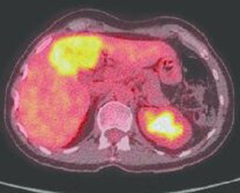
Malignant melanotic nerve sheath tumors (MMNSTs), also known as a melanocytic schwannoma (MS), are a rare type of peripheral nerve sheath tumors including Schwann cells with melanocytic differentiation. Only a few cases of spinal MMNST have been reported in literature. Fluorine-18 fluorodeoxyglucose positron emission tomography/computed tomography ( 18 F-FDG-PET/CT) could be used to detect these lesions. A 70-year-old man with a 6-month history of backache was admitted to our hospital. PET/CT showed a paravertebral soft tissue mass along the spinal nerve at the S2 level with strong FDG uptake, and a nodule with increased FDG uptake in the right lobe of the left liver. A CT-guided biopsy of the S2 lesion was performed. The final diagnosis was spinal MS with hepatic metastasis. The patient received stereotactic body radiation therapy. Herein, we report the PET/CT findings of a case of MS with hepatic metastasis. FDG-PET/CT is helpful in the differential diagnosis of benign and malignant lesions although nonspecific.
Keywords: FDG-PET/CT; MMNST; melanocytic schwannoma.
The Author(s). This is an open access article published by Thieme under the terms of the Creative Commons Attribution License, permitting unrestricted use, distribution, and reproduction so long as the original work is properly cited. ( https://creativecommons.org/licenses/by/4.0/ ).
Conflict of interest statement
Conflict of Interest None declared.
Figures


References
-
- Torres-Mora J, Dry S, Li X, Binder S, Amin M, Folpe A L. Malignant melanotic schwannian tumor: a clinicopathologic, immunohistochemical, and gene expression profiling study of 40 cases, with a proposal for the reclassification of “melanotic schwannoma”. Am J Surg Pathol. 2014;38(01):94–105. - PubMed
-
- Mouchaty H, Conti R, Buccoliero A M, Conti P. Intramedullary melanotic schwannoma of the conus medullaris: a case report. Spinal Cord. 2008;46(10):703–706. - PubMed
-
- Santaguida C, Sabbagh A J, Guiot M C, Del Maestro R F. Aggressive intramedullary melanotic schwannoma: case report. Neurosurgery. 2004;55(06):1430. - PubMed
-
- Lieber B, Han B, Allen J et al.Utility of positron emission tomography in schwannomatosis. J Clin Neurosci. 2016;30:138–140. - PubMed
Publication types
LinkOut - more resources
Full Text Sources