. 2025 Jan 31:16:1547021.
doi: 10.3389/fmicb.2025.1547021. eCollection 2025.

Preparation and application evaluation of monoclonal antibodies against Monkeypox virus A29 protein

Affiliations

Preparation and application evaluation of monoclonal antibodies against Monkeypox virus A29 protein

Ai Xiangjun et al. Front Microbiol. .

Abstract

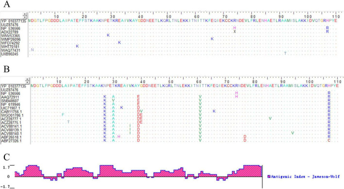
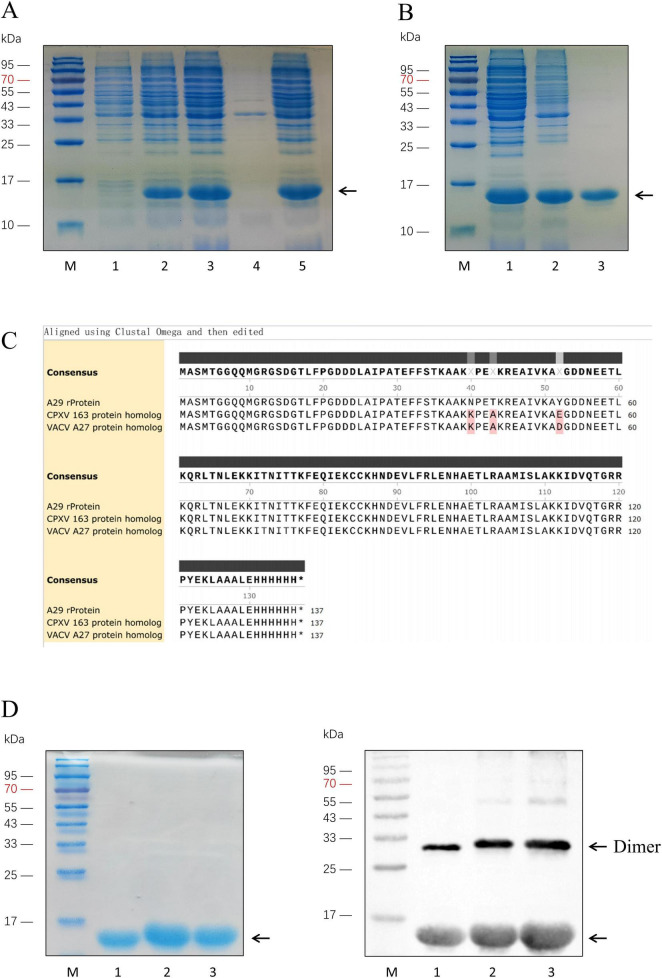
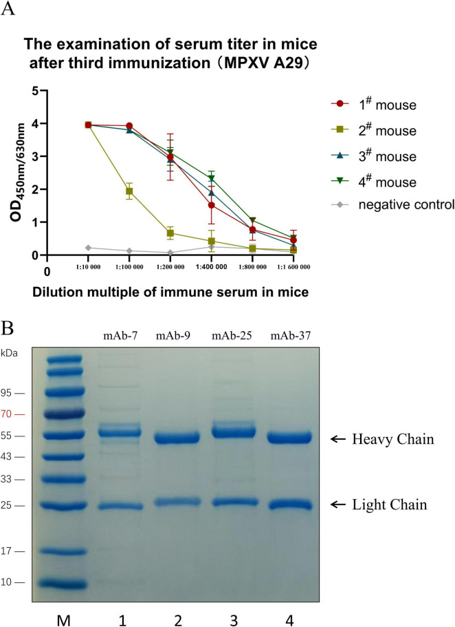
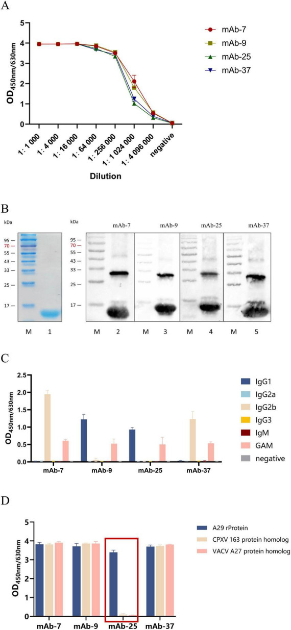
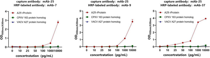
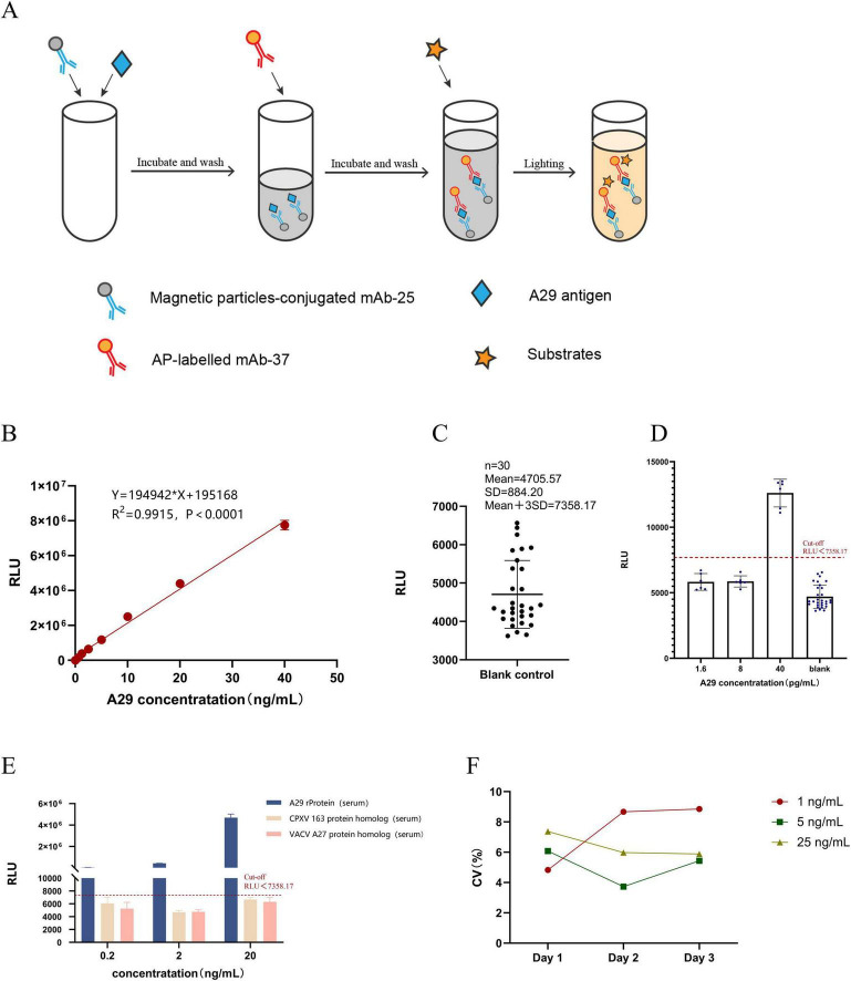
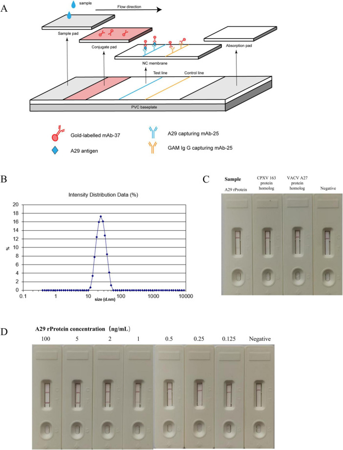
Monkeypox virus (MPXV) is a DNA virus belonging to the Orthopoxvirus genus of the Poxviridae family. It causes symptoms similar to Smallpox virus and is a zoonotic virus with widespread prevalence. Antigen detection is a fast and effective detection method. The MPXV A29 protein not only plays an important role in the virus lifecycle but also serves as a promising target for developing specific antibodies, which have significant potential for application in the diagnosis of MPXV. The coding sequences of the MPXV A29 protein, Cowpox virus (CPXV) 163 protein homolog and Vaccinia virus (VACV) A27 protein homolog were chemically synthesized, and all three recombinant proteins were expressed in Escherichia coli (BL21 Star). Then, the recombinant A29 protein was used as an antigen to immunize BALB/c mice, and a total of 4 monoclonal antibodies against A29 protein were obtained. Using two homologous proteins as reverse screening systems, a specific monoclonal antibody, mAb-25, against the A29 protein was screened. Then, the mAb-25 was used as a coating antibody to pair with other monoclonal antibodies, leading to the identification of a well-matched antibody pair. A chemiluminescence enzyme immunoassay (CLEIA) and immunochromatographic gold assay were subsequently established using the optimal antibody pair. The experimental results indicate that monoclonal antibodies against the A29 protein hold significant potential for application in the diagnosis of MPXV.

Keywords: Monkeypox virus (MPXV); antigen detection; chemiluminescence enzyme immunoassay; colloidal gold immunochromatography; monoclonal antibodies.


Conflict of interest statement

XY, TC, QY, HD, WQ, and LR were employed by the Hunan Xuxiang Biotechnology Co., Ltd. The remaining authors declare that the research was conducted in the absence of any commercial or financial relationships that could be construed as a potential conflict of interest.

Figures

FIGURE 1
FIGURE 1
Bioinformatics analysis of A29 protein. (A) Homology comparison of amino acid sequences of A29 protein from different MPXV subtypes: The colored letters in the figure represent the amino acid composition of the A29 protein, with different colors indicating different types of amino acids. Dots represent amino acids that are identical to those in the first row of the sequence (the recombinant A29 protein expressed in this study), highlighting the differing amino acids. (B) Comparative analysis of the amino acid sequences of MPXV A29 protein and other highly homologous poxvirus proteins: the amino acid sequences in rows 1–3 are from Monkeypox virus, rows 4–6 from Cowpox virus, rows 7–9 from Vaccinia virus, rows 10–11 from Buffalopox virus, rows 12–14 from Camelpox virus, and rows 15–16 from Variola virus. (C) Analysis of antigenic epitopes of A29 protein: the X-axis represents the position of the amino acids in the sequence, the Y-axis represents the antigenicity score for each amino acid position, with higher values indicating stronger antigenicity.
FIGURE 2
FIGURE 2
Expression and purification of A29 protein and its homologous proteins. (A) Identification of recombinant A29 protein expression: Lane M: Protein relative molecular weight markers; Lane 1: Before induction of A29 protein-expressing E. coli strain; Lane 2: After induction of A29 protein-expressing E. coli strain; Lane 3: Suspension of A29 protein-expressing E. coli strain after sonication; Lane 4: Pellet of A29 protein-expressing E. coli strain after sonication; Lane 5: Supernatant of A29 protein-expressing E. coli strain after sonication. (B) Purification of recombinant A29 protein: Lane M: protein relative molecular weight markers; Lane 1: Protein before ammonium sulfate precipitation; Lane 2: Protein after ammonium sulfate precipitation; Lane 3: Protein after His-tag affinity chromatography purification. (C) Comparison of amino acid sequences of the three recombinant proteins: The sequences were derived from the translation of sequencing results of the recombinant plasmids with the T7 promoter from transformed colonies. (D) Identification of three purified proteins: Left: Coomassie Brilliant Blue staining; Right: Western Blot identification; Lane M: Protein relative molecular weight markers; Lane 1: Recombinant A29 protein; Lane 2: CPXV 163 protein homolog; Lane 3: VACV A27 protein homolog.
FIGURE 3
FIGURE 3
Preparation and purification of monoclonal antibodies. (A) Detection of mouse serum titer by indirect ELISA. (B) SDS-PAGE analysis of the purified monoclonal antibody: Lane M: Protein molecular weight markers; Lane 1: mAb-7; Lane 2: mAb-9; Lane 3: mAb-25; Lane 4: mAb-37.
FIGURE 4
FIGURE 4
Characterization of monoclonal antibodies. (A) Determination of monoclonal antibody titer by indirect ELISA. (B) Western Blot analysis of the reactivity of monoclonal antibodies with the protein: Lane M: Protein molecular weight markers; Lane 1: Coomassie Brilliant Blue staining to identify A29 protein; Lanes 2–5: Western Blot identification of A29 protein using mAb-7, mAb-9, mAb-25, and mAb-37 as primary antibodies, respectively. (C) Antibody isotyping. (D) Specificity of the antibody determined by indirect ELISA.
FIGURE 5
FIGURE 5
Screening of antibody pairs by sandwich ELISA. The specific antibody mAb-25 was used as the capture antibody, while the non-specific antibodies mAb-7, mAb-9, and mAb-37 were used as HRP-labeled antibodies to establish a sandwich ELISA.
FIGURE 6
FIGURE 6
Establishment and evaluation of chemiluminescent enzyme immunoassay. (A) Principle of Chemiluminescence Enzyme Immunoassay Method. (B) Construction of the standard curve: Chemiluminescent enzyme immunoassay was used to measure different concentrations of simulated positive samples, and a linear regression analysis was performed between RLU readings and sample concentration. (C) Determination of the cut-off value: Thirty blank samples were measured, and the cut-off value was set as the mean + 3SD. (D) Detection limit determination: The concentration of recombinant A29 protein in simulated positive serum samples was adjusted to different values, and the test was repeated six times. (E) Specificity testing: Simulated positive serum samples (recombinant A29 protein, VACV A27 protein homolog, and CPXV 163 protein homolog) were prepared at three different concentrations (20, 2, and 0.2 ng/mL), with each sample measured in triplicate to determine the RLU value. (F) Stability testing: Simulated positive serum samples (recombinant A29 protein) were prepared at low, medium, and high concentrations. Each sample was measured over three consecutive days, with three replicates per day, to calculate the coefficient of variation.
FIGURE 7
FIGURE 7
Establishment and evaluation of colloidal gold immunochromatographic assay. (A) Schematic diagram of the colloidal gold immunoassay. (B) Measurement of the particle size of gold nanoparticles. (C) Specificity test of colloidal gold test strips: the reactivity of colloidal gold test strips with recombinant A29 protein, VACV A27 protein homolog, and CPXV 163 protein homolog was measured. (D) Sensitivity test of colloidal gold test strips: the reactivity of test strips with samples containing different concentrations of recombinant A29 protein was evaluated.

Similar articles

References

    1. Adegboye O., Alele F., Pak A., Alakunle E., Emeto T., Leggat P., et al. (2024). Monkeypox Outbreak 2022, from a Rare Disease to Global Health Emergence: implications for travellers. Adv. Exp. Med. Biol. 1451 355–368. 10.1007/978-3-031-57165-7_23 - DOI - PubMed
    1. Adegboye O., Gayawan E., James A., Adegboye A., Elfaki F. (2021). Bayesian spatial modelling of Ebola outbreaks in Democratic Republic of Congo through the INLA-SPDE approach. Zoonoses Public Health 68 443–451. 10.1111/zph.12828 - DOI - PubMed
    1. Can G., Perk B., Çitil B. E., Büyüksünetçi Y. T., Anık Ü. (2024). Electrochemical immunoassay platform for human monkeypox virus detection. Anal. Chem. 96 8342–8348. 10.1021/acs.analchem.3c05182 - DOI - PMC - PubMed
    1. Dashprakash M., Venkatesan G., Kumar A., Sankar M., Arya S., Ramakrishnan M. A., et al. (2019). Prokaryotic expression, purification and evaluation of goatpox virus ORF117 protein as a diagnostic antigen in indirect ELISA to detect goatpox. Arch. Virol. 164 1049–1058. 10.1007/s00705-019-04170-8 - DOI - PubMed
    1. Davis I., Payne J. M., Olguin V. L., Sanders M. P., Clements T., Stefan C. P., et al. (2023). Development of a specific MPXV antigen detection immunodiagnostic assay. Front. Microbiol. 14:1243523. 10.3389/fmicb.2023.1243523 - DOI - PMC - PubMed

LinkOut - more resources