. 2024 Dec 30:30:448-464.
eCollection 2024.

Analysis of Smad3 in the modulation of stromal extracellular matrix proteins in corneal scarring after alkali injury

Affiliations

Analysis of Smad3 in the modulation of stromal extracellular matrix proteins in corneal scarring after alkali injury

Suneel Gupta et al. Mol Vis. .

Abstract

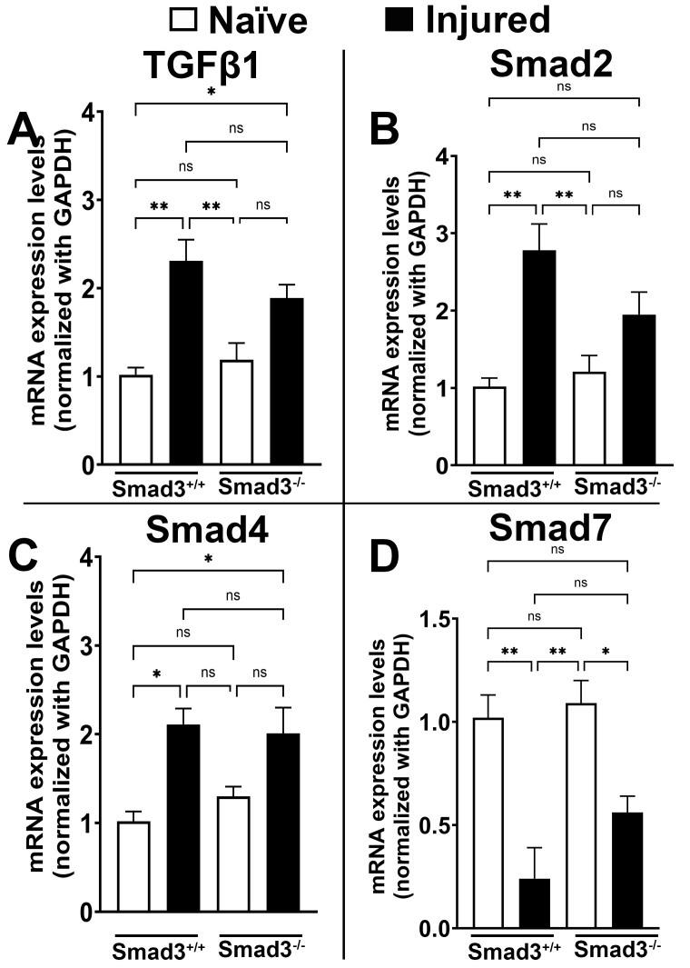
Purpose: During ocular trauma, excessive proliferation and transdifferentiation of corneal stromal fibroblasts cause haze/fibrosis in the cornea. Transforming growth factor β (TGFβ) plays a key role in corneal fibrosis through the Smad signaling pathway. The aberrant activity of TGFβ signaling during ocular trauma (viz. mechanical, infectious, chemical, or surgically altered TGFβ/Smad signaling) leads to regulating the predominant expression of myogenic proteins and the extracellular matrix (ECM). We sought to investigate the functional role of Smad3 in corneal wound repair and stromal ECM assembly using Smad3+/+ wild-type and Smad3-/- deficient mice.

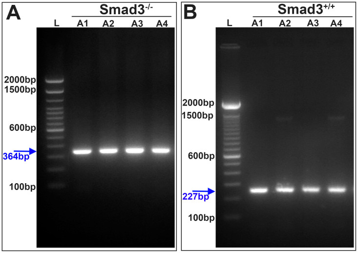
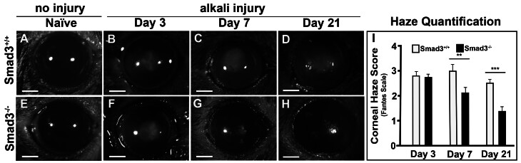
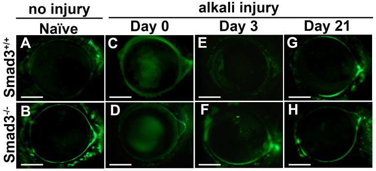
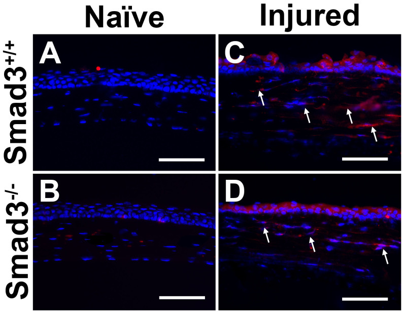
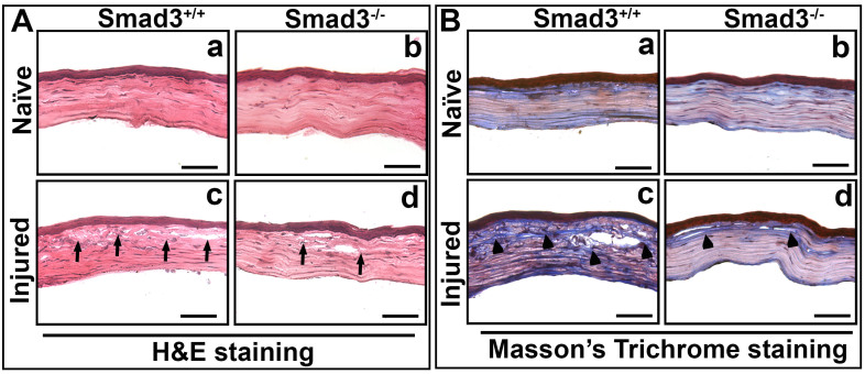
Methods: Corneal injury was introduced with the topical application of an alkali-soaked 2-mm filter disc on the central cornea in the Smad3+/+ (C57BL/6J) and Smad3-/- (129-Smad3tm1Par/J) mouse strains. Slit-lamp and stereo microscopy were used for clinical assessment and corneal haze grading in live animals. Hematoxylin and eosin and Masson's trichrome staining were used to study comparative morphology and collagen level alterations between the groups. Real-time qRT-PCR, western blot, and immunohistochemistry were used to measure changes in profibrotic genes at the mRNA and protein levels.

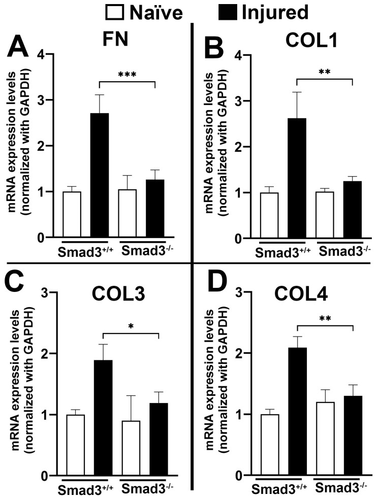
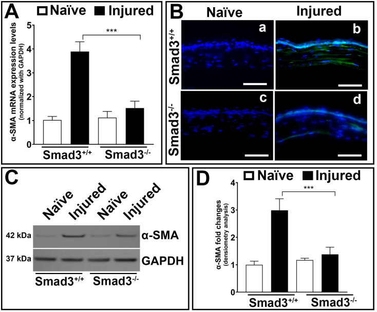
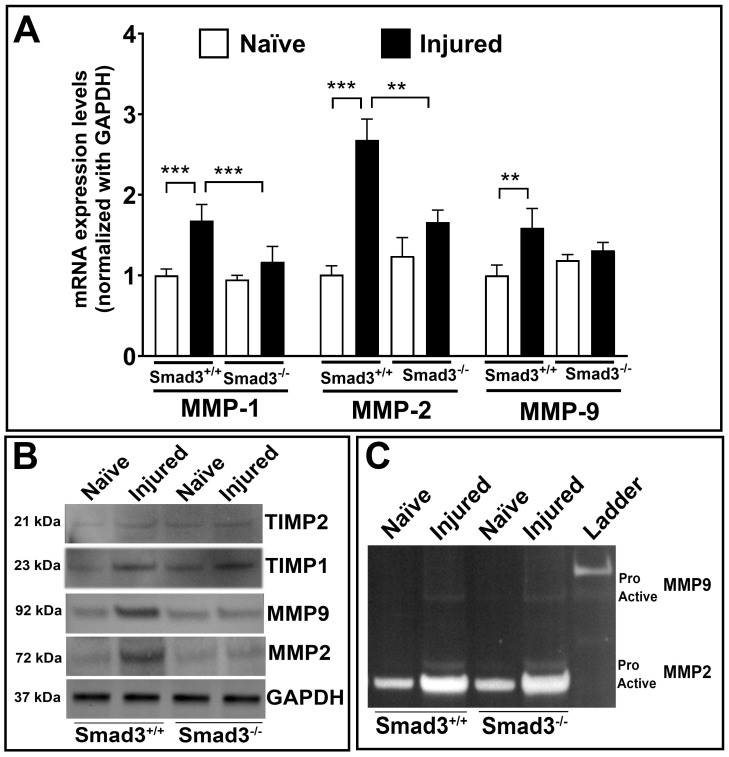
Results: Slit-lamp clinical exams and stereo microscopy detected notably less opaque cornea in the eyes of Smad3-/- compared with Smad3+/+ mice at 3 weeks (p<0.01) in live animals. Corneal tissue sections of Smad3-/- mice showed significantly fewer α-smooth muscle actin-positive cells compared with those of the Smad3+/+ animals (p<0.05). The corneas of the Smad3-/- mice showed significantly lower mRNA levels of pro-fibrotic genes, α-smooth muscle actin, fibronectin, and collagen I (p<0.05, p<0.01, and p<0.001). In addition, the matrix metalloproteinase and tissue inhibitors of metalloproteinase levels were significantly increased (p<0.001) in the corneal tissue during alkali injury in both Smad3+/+ wild-type and Smad3-/- deficient mice.

Conclusions: The significant changes in profibrotic genes and stromal ECM proteins revealed a direct role of Smad3 in stromal ECM proteins and TGFβ/Smad-driven wound healing. Smad3 appears to be an attractive molecular target for limiting abnormal stroma wound healing to treat corneal fibrosis in vivo.


Figures

Figure 1
Figure 1
The 2% agarose gel validates the genotype of Smad3−/− and Smad3+/+ mouse strains. A: The band at 364 bp confirms the Smad3−/− mouse strain in the representative gel image in A1-A4 different mice of Smad3−/− strain genomic DNA samples. B: The band at 227 bp confirms the Smad3+/+ mouse strain in the representative gel image in A1-A3 different mice of Smad3+/+ strain genomic DNA samples. L = 100 bp DNA ladder, bp = base pair.
Figure 2
Figure 2
Smad3 gene deficiency reduced corneal haze in mice after alkali injury. The representative stereomicroscopic mouse corneal tissue images show the A: naïve; B: day 3; C: day 7; and D: day 21 post-alkali haze formation in Smad3+/+ mouse strain corneal tissue. The stereomicroscopic mouse corneal tissue images show the E: naïve; F: day 3; G: day 7; and H: day 21 post-alkali haze formation in Smad3−/− mouse strain corneal tissue. I: The haze quantification graph shows that corneal haze was significantly reduced in Smad3−/− compared with Smad3+/+ mouse corneal tissue (**p<0.01, ***p<0.001, scale bar = 0.5 mm).
Figure 3
Figure 3
Smad3 gene deficiency does not alter the corneal epithelium. A: Smad3+/+ naïve mouse corneal tissue; B: Smad3−/− naïve mouse corneal tissue; C: Smad3+/+ alkali-injured mouse corneal tissue at day 0; D: Smad3−/− alkali-injured mouse corneal tissue at day 0; E: Smad3+/+ alkali-injured mouse corneal tissue at day 3; F: Smad3−/− alkali-injured mouse corneal tissue at day 3; G: Smad3+/+ alkali-injured mouse corneal tissue at day 21; and H: Smad3−/− alkali-injured mouse corneal tissue at day 21. The gross examination shows no alteration in the corneal epithelium of both strains of Smad3+/+ and Smad3−/− naïve and alkali-injured corneas in a time-dependent manner. Scale bar = 0.5 mm.
Figure 4
Figure 4
Smad3 gene deficiency tailored the inflammatory protein expression and impacted the corneal wound healing in mice after alkali injury. Expression of inflammatory protein CD11b expression in A: Smad3+/+ naïve mouse corneal tissue; B: Smad3−/− naïve mouse corneal tissue; C: Smad3+/+ alkali-injured mouse corneal tissue at day 21; and D: Smad3−/− alkali-injured mouse corneal tissue at day 21. The alkali injury increases the CD11b protein expression (arrow) in both strains of Smad3+/+ and Smad3−/− mouse corneal tissue as compared with naïve control mouse corneal tissue, respectively. Scale bar = 200 µm.
Figure 5
Figure 5
Smad3 gene deficiency suppresses the profibrotic gene expression and regulates stromal ECM protein modulation in mice after alkali injury. A: Fibronectin (p<0.001); B: collagen 1 (p<0.01); C: collagen 3 (p<0.05); and D: collagen IV (p<0.01) comparative mRNA expression of Smad3−/− mouse corneal tissue showed significantly reduced expression of profibrotic genes compared with Smad3+/+ mouse corneal tissue after alkali injury. Results are expressed as mean ± SEM, and p<0.05 was considered significant. FN = Fibronectin; COL1 = Collagen 1; COL3 = Collagen 3; and COL4 = Collagen 4.
Figure 6
Figure 6
Smad3 gene deficiency regulates the stromal ECM protein modulation during fibrosis in mice after alkali injury. A: The comparative mRNA expression of α-SMA (fibrosis marker) was significantly downregulated in Smad3−/− mouse corneal tissue compared with Smad3+/+ mouse corneal tissue after alkali injury. B: The immunofluorescence staining of α-SMA was prominently reduced in Smad3−/− mouse corneal tissue compared with Smad3+/+ mouse corneal tissue after alkali injury, whereas no changes were recorded in both Smad3+/+ and Smad3−/− mouse naïve corneal tissue sections. C: The western blot images showed that the α-SMA protein level was notably abridged in Smad3−/− mouse corneal tissue compared with Smad3+/+ mouse corneal tissue after alkali injury, whereas no changes were recorded in both Smad3+/+ and Smad3−/− mouse naïve corneal tissue sections. D: The western blot densitometry analysis graph shows a significant reduction in fibrosis level in Smad3−/− mouse corneas compared with Smad3+/+ mouse corneas. The results are expressed as mean ± SEM, and p<0.05 was considered significant. Scale bar = 200 µm.
Figure 7
Figure 7
Smad3 directly associated with stromal ECM protein modulation impacted the corneal stromal wound healing in mice after alkali injury during active wound healing. A: H&E staining showing the morphological architecture of Smad3+/+ naïve (a); Smad3−/− naïve (b); Smad3+/+ post-alkali injury (c); and Smad3−/−- post-alkali injury (d) corneal tissue sections at day 21. No structural changes were observed in Smad3−/− and Smad3+/+ naïve corneal tissue sections. An increased cellular infiltration and distorted collagen lamellae (arrows) were observed in the corneal stroma of Smad3+/+ and Smad3−/− compared with Smad3−/− mouse corneal tissue sections after alkali injury at day 21. B: Masson’s trichrome staining showed the gross collagen level in Smad3+/+ naïve (a); Smad3−/− naïve (b); Smad3+/+ post-alkali injury (c); and Smad3−/− post-alkali injury (d) corneal tissue sections at day 21. No collagen levels were altered in Smad3−/− and Smad3+/+ naïve corneal tissue sections. Increased collagen deposition was observed in the corneal stroma of Smad3+/+ compared with Smad3−/− mouse corneal tissue sections after alkali injury at day 21, as indicated by increased blue color intensity (arrowhead). Scale bar = 200 μm.
Figure 8
Figure 8
Effects of Smad3 gene deficiency in TGFβ/Smad signaling in mice after alkali injury. A: TGFβ1 (p<0.001); B: Smad2 (p<0.01); C: Smad4 (p<0.05); and D: Smad7 comparative mRNA expression significantly altered in post-alkali injury tissue at day 21 in both Smad3+/+ and Smad3−/− strains as compared with corresponding naïve control mouse corneal tissue. Results are expressed as mean ± SEM, and p<0.05 was considered significant. (*p<0.05, ***p<0.01, and ns = not significant).
Figure 9
Figure 9
Smad3 gene deficiency impacted stromal ECM protein modulation via MMP and TIMP expression during active wound healing in mice after alkali injury. A: The comparative mRNA expressions of MMP1, MMP2, and MMP9 showed significantly reduced expression in Smad3−/− mouse corneal tissue after alkali injury compared with Smad3+/+ mouse corneal tissue after alkali injury. B: The western blot showed similar trends of MMP and TIMP expression in the corneal tissue lysate of Smad3−/− mice compared with Smad3+/+ mouse corneal tissue lysate. C: Gelatin zymography showed the pro and active MMP patterns in the corneal tissue lysate of Smad3−/− mice compared with Smad3+/+ mouse corneal tissue lysate. The results are expressed as mean ± SEM, and p<0.05 was considered significant.
Figure 10
Figure 10
Enhanced Smad3 expression during the post-trauma period played a critical role during wound healing. A: Smad3 level in normal donor human cornea. B: Smad3 level in traumatic human cornea. The Smad3 levels were remarkably increased (arrow) in the post-trauma donor human cornea as compared with the normal donor human cornea. Scale bar = 200 µm.

Similar articles

References

    1. Bukowiecki A, Hos D, Cursiefen C, Eming SA. Wound-Healing Studies in Cornea and Skin: Parallels, Differences and Opportunities. Int J Mol Sci. 2017;18:1257. - PMC - PubMed
    1. Ghafar NA, Jalil NAA, Kamarudin TA. Wound healing of the corneal epithelium: a review. Asian Biomed (Res Rev News) 2021;15:199–212. Res Rev News. - PMC - PubMed
    1. Mohan RR, Gupta S, Kumar R, Sinha NR, Landreneau J, Sinha PR, Tandon A, Chaurasia SS, Hesemann NP. Tissue-targeted and localized AAV5-DCN and AAV5-PEDF combination gene therapy abrogates corneal fibrosis and concurrent neovascularization in rabbit eyes in vivo. Ocul Surf. 2024;32:13–25. - PubMed
    1. Wilson SL, El Haj AJ, Yang Y. Control of scar tissue formation in the cornea: strategies in clinical and corneal tissue engineering. J Funct Biomater. 2012;3:642–87. - PMC - PubMed
    1. Stern JH, Tian Y, Funderburgh J, Pellegrini G, Zhang K, Goldberg JL, Ali RR, Young M, Xie Y, Temple S. Regenerating eye tissues to preserve and restore vision. Cell Stem Cell. 2018;22:834–49. - PMC - PubMed

MeSH terms

LinkOut - more resources