. 2025 Jan 31:16:1528604.
doi: 10.3389/fphar.2025.1528604. eCollection 2025.

Targeted ErbB4 receptor activation prevents D-galactose-induced neuronal senescence via inhibiting ferroptosis pathway

Affiliations

Targeted ErbB4 receptor activation prevents D-galactose-induced neuronal senescence via inhibiting ferroptosis pathway

Ji-Ji Dao et al. Front Pharmacol. .

Abstract

Background: Neuronal senescence is a common pathological feature of various neurodegenerative diseases, with ferroptosis playing a significant role. This study aims to investigate the role of ErbB4 receptor activation in preventing D-Galactose (D-gal)-induced neuronal senescence.

Methods: Mice subjected to D-gal-induced aging were administered a small molecule ErbB4 receptor agonist (E4A), identified via virtual screening, melatonin, or a combination of both. Behavioral assessments were conducted to evaluate therapeutic efficacy in memory and cognitive functions. Immunofluorescence staining, western blot, and biochemical assays were primarily employed to assess changes in both senescence- and ferroptosis-related molecules in mouse hippocampal tissues in response to each treatment. Additionally, mouse hippocampal HT22 neuronal cell cultures were utilized to corroborate the in vivo findings.

Results: The targeted activation of ErbB4 receptor by E4A significantly ameliorated the behavioral deficits induced by D-gal in mice, demonstrating an effect comparable to that of melatonin, a natural inhibitor of in vivo senescence and ferroptosis. Both E4A and melatonin mitigated D-gal-induced aging in hippocampal neurons of mice. This was evidenced by the upregulation of Lamin B1 and the downregulation of P53, P21, P16, GFAP, and Iba-1 expression levels. Moreover, D-gal treatment markedly decreased the protein expression of the ferroptosis inhibitor Nrf2 while augmenting the expression of the ferroptosis promoter TFRC. These alterations were partially reversed by the individual administration of E4A and melatonin. In vitro studies further corroborated that D-gal treatment significantly and concurrently induced the expression of senescence markers and ferroptosis promoters. However, both E4A and melatonin were able to significantly reverse these changes. Additionally, E4A markedly ameliorated Erastin-induced ferroptosis in mouse hippocampal neuronal cells.

Conlusion: Our findings suggest that targeted activation of ErbB4 receptor may be a viable strategy for treating neuronal senescence by inhibiting ferroptosis, thereby offering a potential therapeutic avenue for senescence-associated neurodegenerative diseases.

Keywords: D-galactose; ErbB4 receptor; ferroptosis; hippocampus; neurodegenerative diseases; neuron; senescence; small molecule.


Conflict of interest statement

The authors declare that the research was conducted in the absence of any commercial or financial relationships that could be construed as a potential conflict of interest.

Figures

FIGURE 1
FIGURE 1
The effects of ErbB4 receptor agonist E4A and melatonin on cognitive and spatial memory deficits in D-gal-induced aging mice. (A) Representative swimming trajectories of mice subjected to different treatments in the Morris Water Maze (MWM) spatial probe test. (B) Escape latency across the five experimental groups. (C) Number of target crossings during the MWM probe test. (D) Time spent in the target quadrant during the MWM probe test. (E) Representative heat map showing interaction frequency with novel versus familiar objects. (F) Recognition index in the Novel Object Recognition test among the experimental groups. (G) Spontaneous alternation behaviors in the Y-maze test were assessed. Data were presented as mean ± SD (n = 6–8). Statistical significance was denoted as follows: *p < 0.05, **p < 0.01, ****p < 0.0001.
FIGURE 2
FIGURE 2
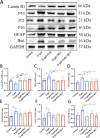
Effect of ErbB4 receptor agonist and melatonin can ameliorate D-gal-induced aging in hippocampus in mice. (A) Western blot analysis of Lamin B1, p53, p21, p16, GFAP, and Iba-1 protein expression levels in the hippocampus of mice. (B–G) Quantification of Lamin B1, p53, p21, p16, GFAP, and Iba-1 protein levels. Data are presented as mean ± SD, with n = 4–5. *p < 0.05, **p < 0.01, ***p < 0.001.
FIGURE 3
FIGURE 3
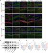
ErbB4 receptor agonist and melatonin inhibit ferroptosis in hippocampus in D-gal-induced aging mice. (A) Immunofluorescence analysis of GPX4-positive cells, SLC7A11-positive cells, TFRC-positive cells in the dentate gyrus (DG) and CA1 regions. Scale bar = 50 μm. (B) Western blot analysis of Nrf2, TFRC, SLC7A11, and GPX4 protein expression levels in the hippocampus of mice. (C–F) Quantification of Nrf2, TFRC, SLC7A11, and GPX4 protein levels. Data are presented as mean ± SD, with n = 4–5. *p < 0.05, **p < 0.01, ***p < 0.001, ****p < 0.0001.
FIGURE 4
FIGURE 4
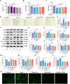
The mitigation of D-gal-induced ferroptosis in HT22 cells by ErbB4 receptor agonist and melatonin. (A) Cell viability post D-gal exposure, evaluated using the CCK-8 assay (n = 5). (B) Cell viability following co-treatment with D-gal and ErbB4 receptor agonist, assessed via the CCK-8 assay (n = 6). (C) Cell viability following co-treatment with D-gal and melatonin, determined by the CCK-8 assay (n = 6). (D) Visualization of cellular senescence through SA-β-gal staining. Scale bar = 100 μm. (E) Quantification of SA-β-gal staining intensities (n = 3). (F) Western blot analysis was conducted to assess the expression levels of senescence-associated markers and ferroptosis-related proteins in HT22 cells. (G–M) Quantification of Nrf2, TFRC, SLC7A11, GPX4, p53, p21, and p16 levels was performed, with normalization to GAPDH (n = 4–7). (N) Intracellular GSH levels (n = 4). (O) Intracellular MDA levels (n = 5). (P) Quantitative analysis of ROS levels was undertaken (n = 4). (Q) Intracellular ROS generation was measured using DCFH-DA, wherein ROS activity was reflected as green fluorescence intensity. Scale bar = 200 μm. Data are presented as mean ± SD. Statistical significance is indicated as follows: *p < 0.05, **p < 0.01, ***p < 0.001, ****p < 0.0001.
FIGURE 5
FIGURE 5
The effects of ErbB4 receptor agonist and melatonin treatment on ferroptosis in Erastin-exposed HT22 cells. (A) Cell viability of HT22 cells post-Erastin exposure was evaluated using the CCK-8 assay (n = 5). (B) Cell viability was evaluated following treatment with Erastin, cotreatment with either E4A or melatonin, or both, utilizing the CCK-8 assay (n = 4). (C) Western blot analysis was conducted to determine the expression levels of senescence-associated markers and ferroptosis-related proteins in HT22 cells. (D–I) Quantitative analysis of Nrf2, TFRC, SLC7A11, GPX4, p21, and p16 levels, normalized to GAPDH (n = 4–7). (J) Intracellular ROS generation was measured using DCFH-DA, with ROS activity indicated by green fluorescence. Scale bar = 200 μm. (K) The quantitative analysis of ROS levels was conducted (n = 4). The data are presented as mean ± SD. Statistical significance is indicated as follows: *p < 0.05, **p < 0.01, ***p < 0.001, ****p < 0.0001.
FIGURE 6
FIGURE 6
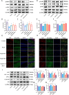
ErbB4 receptor agonist mitigates D-gal-induced hippocampal neuronal aging both in vivo and in vitro through the activation of ErbB4 receptors and modulation of the Akt/Nrf2 signaling pathway. (A) Western blot analysis of pErbB4, ErbB4, pAkt1, and Akt1 protein expression levels in the hippocampus of mice. (B, C) Quantification of pErbB4 and pAkt1 levels (n = 4). (D) Western blot analysis of pErbB4, ErbB4, pAkt1, and Akt1 protein expression levels in D-gal-induced HT22 cells. (E, F) Quantification of pErbB4 and pAkt1 levels (n = 4). (G, H) Immunofluorescence analysis of the Nrf2-positive cells in the DG region (G) and in the cortex region (H). Scale bar = 50 μm. (I) Western blot analysis of Nrf2, SLC7A11, GPX4, Lamin B1 and P21 protein levels after treatment of Nrf2 inhibitor AEM1. (J–N) Quantitative analysis of Nrf2, SLC7A11, GPX4, Lamin B1 and P21 levels (n = 4–7). Data are presented as mean ± SD. Statistical significance is indicated as follows: *p < 0.05, **p < 0.01, ***p < 0.001, ****p < 0.0001.
FIGURE 7
FIGURE 7
Schematic diagram illustrating the effects of targeted activation of ErbB4 receptor on neuronal aging via ferroptosis inhibition. Small molecule agonist (E4A)can activate ErbB4 receptors and regulate the Akt/Nrf2 signaling pathway to achieve neuroprotective effect in D-gal-induced neuronal aging. D-gal treatment increases the accumulation of advanced glycation end products (AGEs) which stimulates the production of reactive oxygen species (ROS) and increase the expression of senescence marker genes P53, P16, and P21. E4A can promote Akt1 phosphorylation and promote Nrf2 entrance into the nucleus, in which it involves in regulating the expression of ferroptosis suppressor gene, such as SLC7A11 and GPX4 to attenuate lipid peroxidation, which can inhibit cell ferroptosis and neuronal aging. The diagram was created with MedPeer (www.medpeer.cn).

References

    1. Ahsan A., Hiniker S. M., Davis M. A., Lawrence T. S., Nyati M. K. (2009). Role of cell cycle in epidermal growth factor receptor inhibitor-mediated radiosensitization. Cancer Res. 69 (12), 5108–5114. 10.1158/0008-5472.CAN-09-0466 - DOI - PMC - PubMed
    1. Ashraf A., Jeandriens J., Parkes H. G., So P. W. (2020). Iron dyshomeostasis, lipid peroxidation and perturbed expression of cystine/glutamate antiporter in Alzheimer's disease: evidence of ferroptosis. Redox Biol. 32, 101494. 10.1016/j.redox.2020.101494 - DOI - PMC - PubMed
    1. Becker N. V., Scott J. W., Moniz M. H., Carlton E. F., Ayanian J. Z. (2022). Association of chronic disease with patient financial outcomes among commercially insured adults. JAMA Intern Med. 182 (10), 1044–1051. 10.1001/jamainternmed.2022.3687 - DOI - PMC - PubMed
    1. Bellinger F. P., Bellinger M. T., Seale L. A., Takemoto A. S., Raman A. V., Miki T., et al. (2011). Glutathione peroxidase 4 is associated with neuromelanin in substantia nigra and dystrophic axons in putamen of Parkinson's brain. Mol. Neurodegener. 6 (1), 8. 10.1186/1750-1326-6-8 - DOI - PMC - PubMed
    1. Bishop N. A., Lu T., Yankner B. A. (2010). Neural mechanisms of ageing and cognitive decline. Nature 464 (7288), 529–535. 10.1038/nature08983 - DOI - PMC - PubMed

LinkOut - more resources