. 2024 Sep 26;5(4):e00114.
doi: 10.1227/neuprac.0000000000000114. eCollection 2024 Dec.

BrainPath Tubular Retractor System for Subcortical Hemorrhagic Vascular Lesions: A Case Series of Technique and Outcomes

Affiliations

BrainPath Tubular Retractor System for Subcortical Hemorrhagic Vascular Lesions: A Case Series of Technique and Outcomes

Leonard H Verhey et al. Neurosurg Pract. .

Abstract

Background and objectives: Hemorrhagic subcortical vascular lesions such as cavernous malformations (CM) and arteriovenous malformations (AVM) can be neurologically devastating. Conventional open surgical resection is often associated with additional morbidity. The BrainPath® (NICO Corp.) transsulcal tubular retractor system offers a less-invasive corridor to deep-seated lesions. Our objective was to describe a single-center experience with the resection of subcortical hemorrhagic vascular lesions in adult and pediatric patients using the BrainPath® system.

Methods: The departmental database was queried for patients who underwent resection of a hemorrhagic CM, AVM, or cerebral aneurysm through the BrainPath® tubular retractor system between January 2017 and September 2021. All patients underwent either postoperative MRI (for patients with CM) or digital subtraction angiography (for patients with AVM or aneurysm). Demographic and clinical characteristics, preoperative and postoperative imaging features, operative details, and surgical and clinical outcomes were extracted through a retrospective review of the medical records.

Results: Fourteen patients (mean [SD] age 32.3 [23.9] years; 7 (50%) female) underwent BrainPath®-based resection of a deeply seated CM (n = 7), AVM (n = 6), or ruptured cerebral aneurysm (n = 1). The mean maximal lesion diameter was 21.5 (12.6) mm. The mean operative time was 134 (53) minutes. Residual lesion was present in 2 patients, both of which underwent repeat BrainPath®-assisted surgery for complete resection. All lesions were completely resected or obliterated on postoperative MRI or digital subtraction angiography. At a mean follow-up of 4.1 (1.1) years, the median modified Rankin Scale score was 1 (range 0-6).

Conclusion: In a well-selected cohort, we show the effective use of BrainPath® tubular retractors for resection or obliteration of subcortical hemorrhagic vascular lesions. This report further exemplifies the expanded role of the endoport system beyond that of intracerebral hemorrhage and tumor. Further study will elucidate the impact of this less-invasive brain retraction technique on clinical outcome in patients with vascular lesions.

Keywords: Arteriovenous malformation; BrainPath; Case series; MIPS; Subcortical cavernous malformation.


Conflict of interest statement

The authors have no personal, financial, or institutional interest in any of the drugs, materials, or devices described in this article.

Figures

FIGURE 1.
FIGURE 1.
A 73-year-old man with ruptured right anterior choroidal artery aneurysm. A-D, Axial head CT demonstrating intraventricular hemorrhage with acute ventriculomegaly and right temporal subarachnoid hemorrhage. E, Preoperative lateral DSA of the right internal carotid injection demonstrating a 2-mm anterior choroidal artery aneurysm. F, Initial postoperative DSA demonstrating residual aneurysmal filling. G and H, Axial and coronal CT angiogram of the head after taken back to the operating room demonstrating complete obliteration of the aneurysm. CT, computed tomography; DSA, digital subtraction angiography.
FIGURE 2.
FIGURE 2.
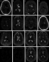
A 4-year-old girl with a hemorrhagic left thalamic cavernous malformation. A, Axial head CT, B and C, axial and coronal T2-weighted, and D, axial gradient echo images at the time of clinical presentation. E, Postoperative axial T2-weighted MRI demonstrating resection cavity. F and G, Routine surveillance axial and coronal T2-weighted MRI at 1 month after surgery demonstrating recurrent hemorrhage and mass effect. H and I, Axial head CT and T2-weighted MRI 10 months after initial craniotomy when patient returned with acute headache and vomiting, demonstrating acute left thalamic hemorrhage with perilesional edema and mass effect. J-L, Axial T2-weighted MRI obtained postoperatively (ie, after second resection) demonstrating interval debulking but residual cavernoma in the left thalamic region. The patient was taken back to the operating room 5 days later for complete resection of residual cavernoma, as demonstrated in intraoperative axial T2-weighted images in M and N. O and P, Last available surveillance scan obtained 18 months after initial presentation continues to demonstrate no residual lesion on axial T2-weighted MRI. The patient's guardians consented to the publishing of these deidentified radiographic images. CT, computed tomography.
FIGURE 3.
FIGURE 3.
A 28-eight-year old woman with a hemorrhagic Spetzler-Martin grade III arteriovenous malformation. A, Axial head CT at the time of presentation with sudden onset headache, nausea, and vomiting demonstrates right thalamic region intracerebral hemorrhage with associated intraventricular hemorrhage and mass effect. B and F, Preoperative lateral and A-P digital subtraction angiography demonstrates an AVM arising from the right P3 branch with a nidus measuring 11 × 9 mm and with associated early deep venous drainage into the vein of Galen complex. C and G, Lateral and A-P projections after Onyx embolization of the nidus through 2 pedicle feeders by a right posterior cerebral artery microcatheterization. E, Axial head CT obtained postoperatively demonstrating a right occipital trajectory to the AVM. D and H, Lateral and A-P projections obtained after craniotomy for AVM resection demonstrating no residual early venous drainage and no residual AVM nidus. AVM, arteriovenous malformation; CT, computed tomography.
FIGURE 4.
FIGURE 4.
A 36-year-old man with hemorrhagic cavernoma in the right thalamus and midbrain. A and B, Axial head CT obtained at the time of presentation with acute left hemiparesis and dysarthria, demonstrating an intracerebral hemorrhage in the right thalamus and extending into the right cerebral peduncle with surrounding edema and regional mass effect. C and D, Axial T2-weighted MRI through the thalamus and midbrain demonstrating a mixed intensity well-circumscribed lesion with intralesional fluid-fluid levels and surrounding edema. E and F, Axial head CT obtained after left frontal craniotomy demonstrating interval resection of hemorrhagic lesion with an external ventricular drain after the surgical trajectory. G and H, Axial T2-weighted MRI obtained approximately 4 months postoperatively demonstrating full resection of the cavernoma with some residual hemosiderin signal change within the thalamus and midbrain. CT, computed tomography.

References

    1. Murthy SB, Merkler AE, Omran SS, et al. Outcomes after intracerebral hemorrhage from arteriovenous malformations. Neurology. 2017;88(20):1882-1888. - PMC - PubMed
    1. Mouchtouris N, Chalouhi N, Chitale A, et al. Management of cerebral cavernous malformations: from diagnosis to treatment. ScientificWorldJournal. 2015;2015:808314. - PMC - PubMed
    1. Zetterling M, Elf K, Semnic R, Latini F, Engström ER. Time course of neurological deficits after surgery for primary brain tumours. Acta Neurochir (Wien). 2020;162(12):3005-3018. - PMC - PubMed
    1. Nico Brainpath. NICO BrainPath Product Information. NICO Inc; 2023. https://niconeuro.com/wp-content/uploads/2020/06/3_LIT208RevH_BrainPath-.... Accessed October 10, 2023.
    1. Mansour S, Echeverry N, Shapiro S, Snelling B. The use of Brainpath tubular retractors in the management of deep brain lesions: a review of current studies. World Neurosurg. 2020;134:155-163. - PubMed

LinkOut - more resources