. 2025 Jan 27:16:1514010.
doi: 10.3389/fendo.2025.1514010. eCollection 2025.

Acupuncture mediates the "gut-testis axis" to improve asthenozoospermia

Affiliations

Acupuncture mediates the "gut-testis axis" to improve asthenozoospermia

Jianheng Hao et al. Front Endocrinol (Lausanne). .

Abstract

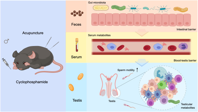
Background: Asthenozoospermia is a common cause of male infertility. Studies have shown that sperm quality and motility are affected by the gut-testis axis that can regulate testicular metabolism and function through the gut microbiota and its metabolites. Acupuncture is an important modality of complementary and alternative medicine. It can improve sperm motility, but it remains unclear whether acupuncture can enhance sperm vitality by influencing the gut-testis axis.

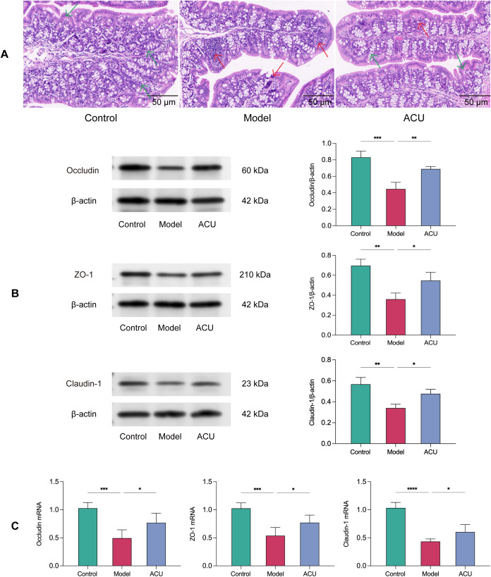
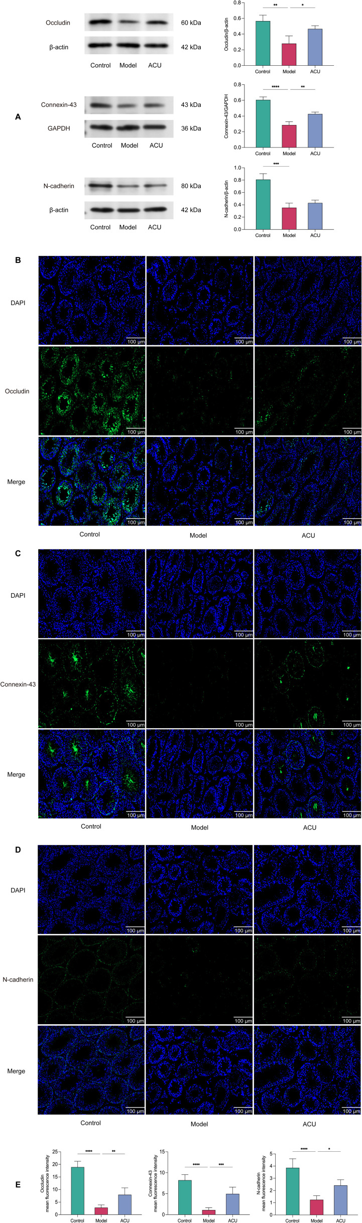
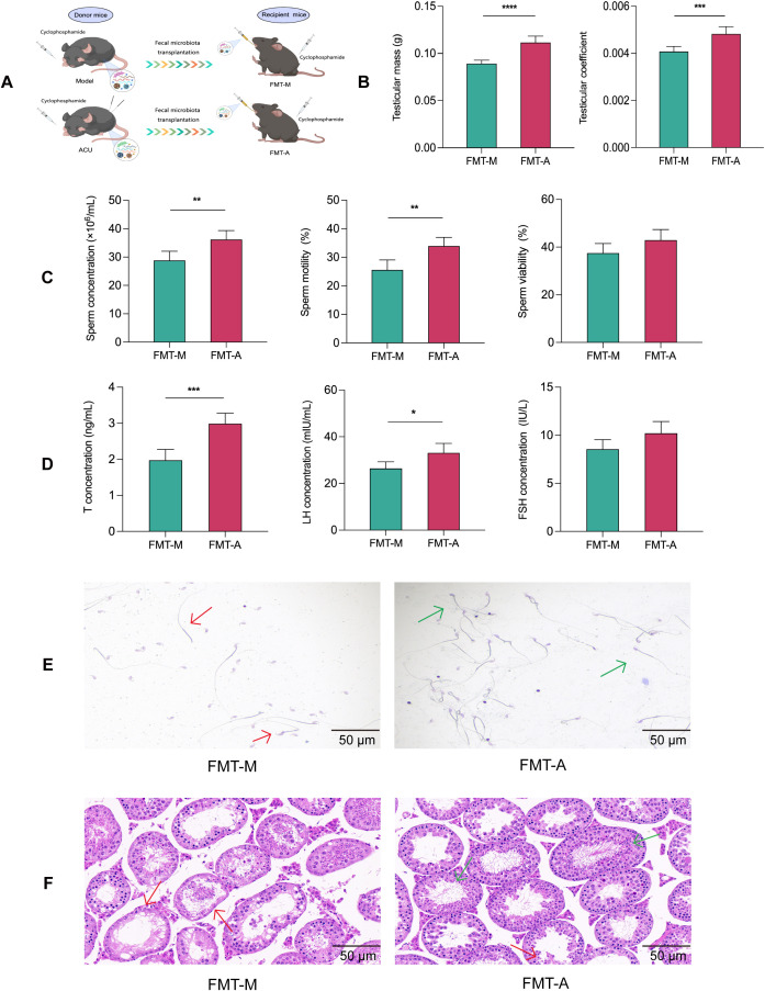
Methods: In this study, sperm quality, testicular pathology, and serum hormone levels were assessed using a cyclophosphamide-induced mouse model. Real-time PCR, a western blot analysis, and immunofluorescence techniques were used to assess the effects of acupuncture on the gut barrier and blood-testis barrier functions. In addition, gut microbiome and metabolomics were used to study the impact of acupuncture on the gut microbiota structure, serum, and testicular metabolites in asthenozoospermic mice. Further validation was obtained by performing a fecal microbiota transplantation (FMT).

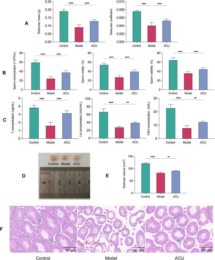
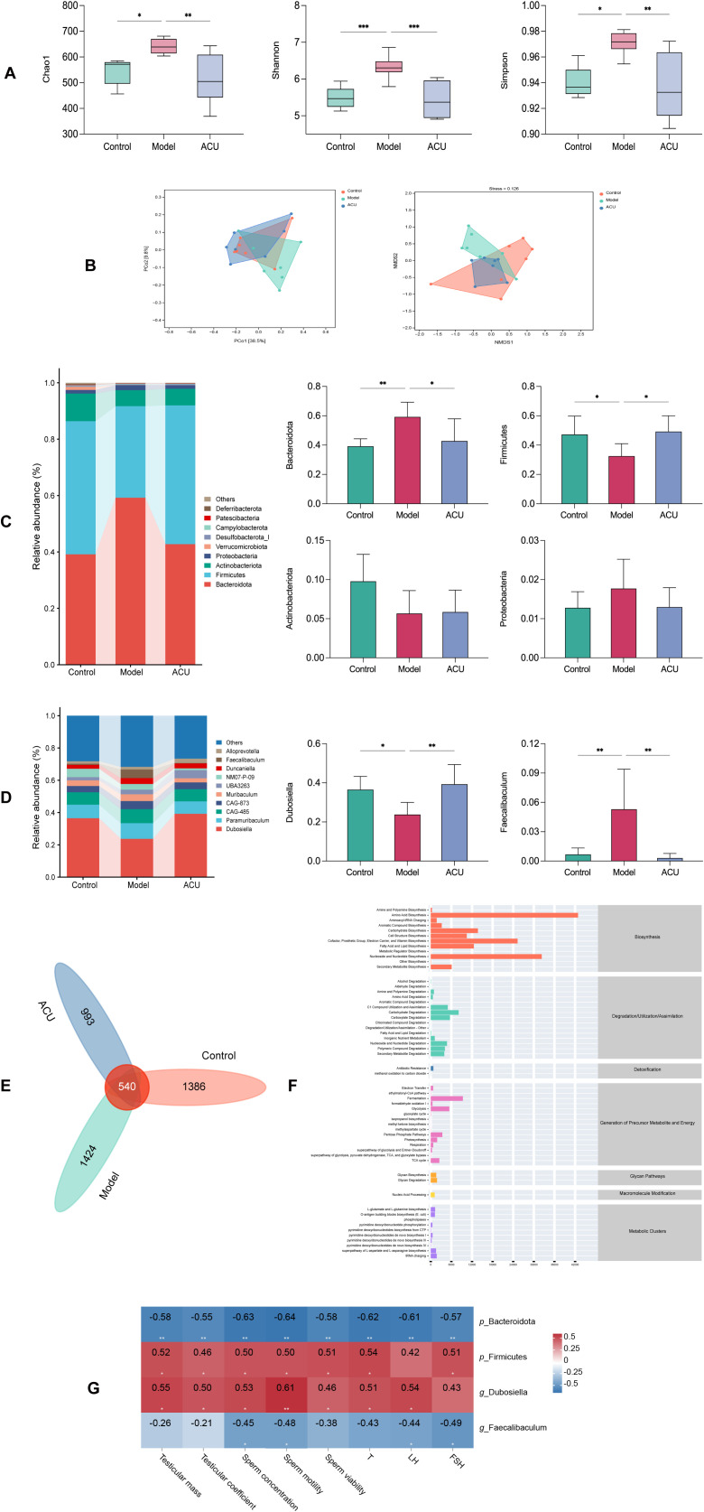
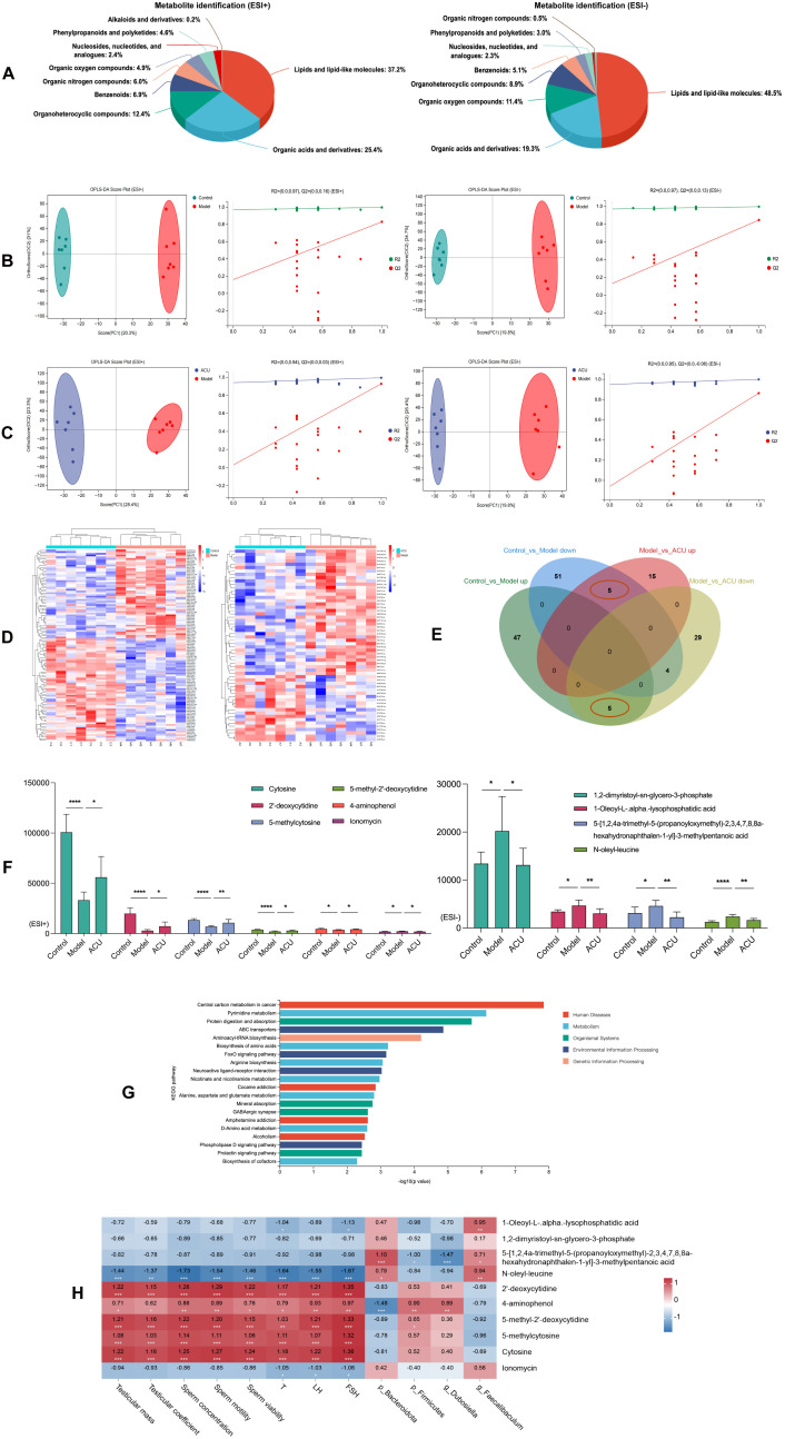
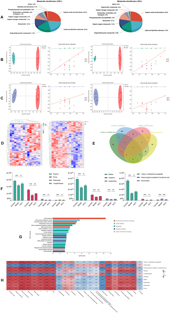
Results: Acupuncture improved the sperm quality; ameliorated testicular pathology; increased serum testosterone (T), follicle-stimulating hormone (FSH), and luteinizing hormone (LH) levels; and repaired gut and blood-testis barrier damage in asthenozoospermic mice. The abundances of Bacteroidota, Firmicutes, Faecalibaculum, and Dubosiella were associated with sperm motility, as shown by a gut microbiome analysis. Serum metabolomics revealed that differentially expressed metabolites (DEMs), such as cytosine and N-oleyl-leucine, were closely related to sperm motility. Testicular metabolomics analysis revealed DEMs, such as 5-fluorouridine and 1-acetylimidazole, were also associated with sperm motility. Furthermore, reproductive function improvements in asthenozoospermic mice through acupuncture were achieved via an FMT.

Conclusion: Acupuncture may alleviate asthenozoospermia symptoms by modulating the gut-testis axis and repairing the gut-testis barrier.

Keywords: acupuncture; asthenozoospermia; gut microbiota; gut-testis axis; metabolomics.


Conflict of interest statement

The authors declare that the research was conducted in the absence of any commercial or financial relationships that could be construed as a potential conflict of interest.

Figures

Figure 1
Figure 1
Acupuncture improved the testicular function and sperm quality in the asthenozoospermic mice. (A) Comparison of testicular mass and testicular coefficient of each group (n=14). (B) Comparison of sperm parameters of each group (n=14). (C) Comparison of serum T, FSH, and LH concentrations of each group (n=7). (D) Comparison of visual observation of testicles of each group. (E) Comparison of testicular volume of each group (n=7). (F) Comparison of testicular histopathological morphology of each group (n=7). Green arrows indicate seminiferous tubules with normal morphology and structure, red arrows indicate tubules with uneven wall thickness and dilated lumens, and blue arrows indicate tubules with improved morphology and structure. Data are presented as mean ± SD. **P < 0.01, ****P < 0.0001.
Figure 2
Figure 2
Acupuncture regulated the gut microbiota structure in the asthenozoospermic mice. (A) Comparison of α-diversity indices of each group. (B) Comparison of β-diversity of each group. (C) Comparison of differences at the phylum level of each group. (D) Comparison of differences at the genus level of each group. (E) Venn diagram of shared species. (F) Predicted metabolic pathways of shared species. (G) Spearman correlation analysis. Data are presented as mean ± SD. *P < 0.05, **P < 0.01, ***P < 0.001.
Figure 3
Figure 3
Acupuncture improved intestinal barrier damage in the asthenozoospermic mice. (A) Comparison of pathological morphology of colon tissues of each group (n=7). (B) Comparison of Occludin, ZO-1, and Claudin-1 protein expression levels in colonic tissues of each group (n=3). (C) Comparison of Occludin, ZO-1, and Claudin-1 mRNA expression levels in colonic tissues of each group (n=5). Note: The green arrows indicate morphologically normal colonic tissue, while the red arrows indicate colonic tissue with disrupted intestinal barrier and inflammatory infiltration. Data are presented as mean ± SD. *P < 0.05, **P < 0.01, ***P < 0.001, ****P < 0.0001.
Figure 4
Figure 4
Acupuncture regulated serum metabolic disorders in the asthenozoospermic mice. (A) Serum metabolite identification and analysis in ESI+ and ESI- Modes. (B) OPLS-DA analysis and permutation test between control and model groups in ESI+ and ESI- Modes. (C) OPLS-DA analysis and permutation test between model and ACU groups in ESI+ and ESI- Modes. (D) Clustering analysis of metabolites among groups in combined ESI+ and ESI- modes. (E) Venn diagram of intersecting differential metabolites in combined ESI+ and ESI- modes. (F) Comparison of differential metabolite expression between ESI+ and ESI- modes. (G) KEGG Enrichment analysis. (H) Spearman correlation analysis. *P < 0.05, **P < 0.01, ***P < 0.001, ****P < 0.0001.
Figure 5
Figure 5
Acupuncture improved BTB damage in the asthenozoospermic mice. (A) Comparison of Occludin, Connexin-43, and N-cadherin protein expression levels in the testicular tissues of each group (n=3). (B-E) Immunofluorescence comparison of Occludin, Connexin-43, and N-cadherin in the testicular tissues of each group (n=3). Data are presented as mean ± SD. *P < 0.05, **P < 0.01, ***P < 0.001, ****P < 0.0001.
Figure 6
Figure 6
Acupuncture regulated testicular metabolic disorders in the asthenozoospermic mice. (A) Testicular metabolite identification and analysis in ESI+ and ESI- Modes. (B) OPLS-DA analysis and permutation tests between the control and model groups under ESI+ and ESI- modes. (C) OPLS-DA analysis and permutation tests between the model and ACU groups under ESI+ and ESI- modes. (D) Clustering analysis of testicular DEMs between groups under the combined ESI+ and ESI- modes. (E) Venn diagram of intersecting DEMs under the combined ESI+ and ESI- modes. (F) Comparison of expression levels of the top 10 DEMs by VIP values. (G) KEGG enrichment analysis. (H) Spearman correlation analysis. *P < 0.05, **P < 0.01, ***P < 0.001, ****P < 0.0001.
Figure 7
Figure 7
FMT improved reproductive function in the asthenozoospermic mice. (A) Scheme of FMT experiment. (B) Comparison of testicular mass and testicular coefficient of each group (n=6). (C) Comparison of sperm parameters of each group (n=3). (D) Comparison of serum T, FSH, and LH concentrations of each group (n=6). (E) Comparison of sperm pathological morphology of each group (n=3). (F) Comparison of testicular histopathological morphology of each group (n=6). In (E) red arrows indicate abnormal sperm morphology, such as headless sperm and sperm with abnormal tail bending or folding, while green arrows indicate normal sperm morphology. In (F) red arrows indicate seminiferous tubules with uneven wall thickness and dilated lumens, while green arrows indicate seminiferous tubules with improved structural integrity. Data are presented as mean ± SD. *P < 0.05, **P < 0.01, ***P < 0.001, ****P < 0.0001.
Figure 8
Figure 8
Graphical abstract. Acupuncture modulates gut microbiota composition and structure in asthenozoospermic mice, enhancing intestinal barrier integrity and correcting serum metabolic imbalances. This leads to restoration of BTB function and regulation of testicular metabolism, ultimately improving sperm motility.

Similar articles

References

    1. Agarwal A, Baskaran S, Parekh N, Cho CL, Henkel R, Vij S, et al. . Male infertility. Lancet. (2021) 397:319–33. doi: 10.1016/S0140-6736(20)32667-2 - DOI - PubMed
    1. World Health Organization . WHO laboratory manual for the examination and processing of human semen. 5th ed. Geneva: World Health Organization; (2021), ISBN: ISBN: 9789241547789.
    1. Heidary Z, Saliminejad K, Zaki-Dizaji M, Khorram Khorshid HR. Genetic aspects of idiopathic asthenozoospermia as a cause of male infertility. Hum Fertil (Camb). (2020) 23:83–92. doi: 10.1080/14647273.2018.1504325 - DOI - PubMed
    1. Ribeiro JC, Nogueira-Ferreira R, Amado F, Alves MG, Ferreira R, Oliveira PF. Exploring the role of oxidative stress in sperm motility: a proteomic network approach. Antioxid Redox Signal. (2022) 37:501–20. doi: 10.1089/ars.2021.0241 - DOI - PubMed
    1. Nowicka-Bauer K, Lepczynski A, Ozgo M, Kamieniczna M, Fraczek M, Stanski L, et al. . Sperm mitochondrial dysfunction and oxidative stress as possible reasons for isolated asthenozoospermia. J Physiol Pharmacol. (2018) 69:403–17. doi: 10.26402/jpp.2018.3.05 - DOI - PubMed

LinkOut - more resources