. 2023 Mar 6;7(1):90-98.
doi: 10.1016/j.livres.2023.02.004. eCollection 2023 Mar.

Granulocyte colony-stimulating factor reduces biliary fibrosis and ductular reaction in a mouse model of chronic cholestasis

Affiliations

Granulocyte colony-stimulating factor reduces biliary fibrosis and ductular reaction in a mouse model of chronic cholestasis

Trinh Van Le et al. Liver Res. .

Abstract

Background: Biliary atresia is a rare congenital bile duct disease that is the leading cause of liver fibrosis in neonates. Granulocyte colony-stimulating factor (GCSF) is a potential therapy for hepatocellular diseases, but data on GCSF for cholestatic conditions remain limited.

Materials and methods: The current study examines the role of GCSF in improving bile duct obstruction in mice. Two doses were administered: 10.0 μg/kg/day and 61.5 μg/kg/day, which is the animal equivalent dose of 5.0 μg/kg in humans. Seven days (D7) after bile duct ligation (BDL), Swiss mice were treated with phosphate buffered saline or GCSF for 5 days. The intrahepatic adaptive response of BDL mice was evaluated on postsurgical days D12, D19, and D26.

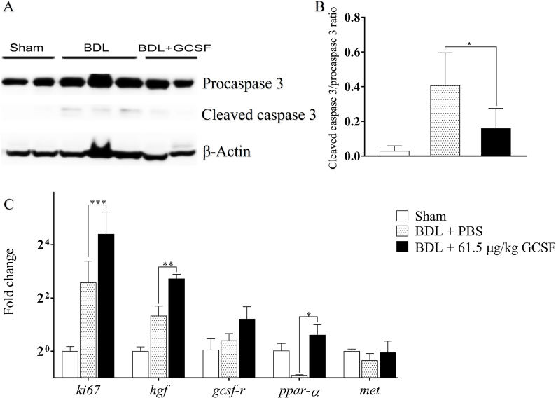
Results: Treatment with 61.5 μg/kg of GCSF resulted in a significant increase in circulating leukocytes and neutrophils on D12. Amelioration of liver injury, as shown by reduced aspartate aminotransferase levels, increased albumin levels and survival rate, as well as reduced intrahepatic inflammation and hepatic myeloperoxidase expression, downregulated ductular proliferation, periportal fibroblast activation, and fibrosis, enhanced expressions of hepatocyte growth factor, peroxisome proliferator-activated receptor-alpha, and ki67, and suppressed expression of cleaved caspase-3 protein, was noted after treatment with 61.5 μg/kg of GCSF. Additionally, GCSF treatment was associated with an increased number of intrahepatic cd3-Sca1+c-Kit+ bone marrow cells.

Conclusions: Treatment with 61.5 μg/kg of GCSF resulted in liver regeneration and survival in BDL mice was seen, suggesting its potential use for human liver diseases.

Keywords: Bile duct ligation (BDL); Biliary fibrosis; Ductular reaction; Granulocyte colony-stimulating factor (GCSF); Hepatic stellate cell (HSC).


Conflict of interest statement

The authors declare that they have no conflict of interest.

Figures

Fig. 1
Fig. 1
Granulocyte colony-stimulating factor (GCSF) improves survival, body weight, and circulating leukocytes in mice that underwent bile duct ligation (BDL) in a dose-dependent manner.(A) The 26-day Kaplan–Meier survival curve of BDL mice treated with phosphate buffered saline (PBS) and 10.0 μg/kg or 61.5 μg/kg of GCSF (n = 10 per group). (B) Change in the body weight of BDL mice treated with PBS and 10.0 μg/kg or 61.5 μg/kg of GCSF (n = 6 per group for each time-point). (C) Circulating leukocyte count and (D) neutrophil percentage on 12 days after surgery (D12), D19, and D26 in BDL mice (n = 6 per group for each time-point). Data are presented as mean ± standard deviation (SD). ∗P < 0.05, ∗∗P < 0.01, ∗∗∗∗P < 0.0001.
Fig. 2
Fig. 2
Effects of GCSF on liver enzymes, hepatic biliary infarction, and inflammation. The serum levels of (A) AST and (B) albumin in BDL mice treated with PBS, 10.0 μg/kg of GCSF, and 61.5 μg/kg of GCSF on 12 days after surgery (D12), D19, and D26. (C) Shown are representative hematoxylin and eosin (H&E) liver micrographs of BDL mice treated with PBS, 10.0 μg/kg of GCSF, and 61.5 μg/kg of GCSF on D26. Green arrows represent leukocyte infiltration. Scale bar = 100 μm. (D) HAI scores of BDL mice treated with PBS, 10.0 μg/kg of GCSF, and 61.5 μg/kg of GCSF on D19 and D26. (E) Representative Sirius-stained liver micrographs (green arrows represent Sirius-positive areas at the periportal spaces of BDL mice treated with PBS, 10.0 μg/kg of GCSF, and 61.5 μg/kg of GCSF on D26. Scale bar = 100 μm. (F) Statistics of Sirius red positive areas. Data are presented as mean ± standard deviation (SD), ∗P < 0.05, ∗∗P < 0.01, ∗∗∗P < 0.001, ∗∗∗∗P < 0.0001 (n = 6 per group for each time-point). Abbreviations: AST, aspartate aminotransferase; BDL, bile duct ligation; GCSF, granulocyte colony-stimulating factor; HAI, histology activity index; PBS, phosphate buffered saline.
Fig. 3
Fig. 3
GCSF reduces liver fibrosis in BDL mice. (A) Immunostaining liver of sham and BDL mice treated with PBS or 61.5 μg/kg GCSF on 26 days after surgery (D26). Scale bar = 50 μm. (B) Quantitation of α-sma positive area in the liver. (C) Western blotting was performed to detect the protein expression of α-sma in livers of mice on D26. (D) Fibrosis-related gene expressions of tgf-β1, α-sma, and mmp9 in the liver of sham and BDL treated with PBS or high-dose GCSF on D26. Data are presented as mean ± standard deviation (SD), ∗P < 0.05, ∗∗P < 0.01 (n = 6 per group). Abbreviations: BDL, bile duct ligation; GCSF, granulocyte colony-stimulating factor; IHC, immunohistochemistry; mmp9, matrix metalloprotease 9; PBS, phosphate buffered saline; α-sma, alpha-smooth muscle actin; tfg-β1, transforming growth factor-beta 1.
Fig. 4
Fig. 4
GCSF enhances hepatic proliferative gene expression and reduces hepatic apoptosis in BDL mice. (A) Western blotting for procaspase 3 and cleaved caspase 3 in the liver. (B) Graph showing the ratio of cleaved caspase 3 and procaspase 3. (C) Real-time PCR results of the expressions of ki67, hgf, gcsf-r, ppar-α, and met on 26 days after surgery (D26) in mice. Data are presented as mean ± standard deviation (SD), ∗P < 0.05, ∗∗P < 0.01, ∗∗∗P < 0.001 (n = 6 per group). Abbreviations: BDL, bile duct ligation; GCSF, granulocyte colony-stimulating factor; hgf, hepatocyte growth factor; PBS, phosphate buffered saline; PCR, polymerase chain reaction; ppar-α, peroxisome proliferator-activated receptor-alpha.
Fig. 5
Fig. 5
GCSF downregulates ductal proliferation in BDL mice. (A) Representative micrographs of ck7-staining on 26 days after surgery (D26) in sham mice and BDL mice treated with PBS or 61.5 μg/kg of GCSF (yellow arrows point to ck7-positive periportal ductular reaction). Scale bar = 100 μm. (B) Average number of ck7 positive bile ducts. (C) The protein expression of ck7 was detected by Western blotting. (D) Quantification of ck7 on D26 in BDL mice treated with PBS or GCSF. (E) Real-time PCR was used to analyze the ck19 expression level. Data are presented as mean ± standard deviation (SD), ∗P < 0.05, ∗∗∗P < 0.001, ∗∗∗∗P < 0.0001 (n = 6 per group). Abbreviations: BDL, bile duct ligation; ck, cytokeratin; GCSF, granulocyte colony-stimulating factor; HKP, house keeping protein; PBS, phosphate buffered saline; PCR, polymerase chain reaction.
Fig. 6
Fig. 6
GCSF enhances hepatic bone marrow stem cell mobilization. Flow cytometry analysis of bone marrow stem cells (cd3-Sca1+c-Kit+) in cd3 liver cell population on 12 days after surgery (D12) in (A) sham mice and BDL mice treated with (B) PBS or (C) 61.5 μg/kg of GCSF. (D) Statistics comparing the three experimental groups. Data are presented as mean ± standard deviation (SD), ∗P < 0.05, ∗∗P < 0.01, n = 4 per group. Abbreviations: BDL, bile duct ligation; GCSF, granulocyte colony-stimulating factor; PBS, phosphate buffered saline.
Supplementary Fig. 1
Liver pathology of bile duct ligation (BDL) mouse models. The sham and BDL mice model indices changed in (A) body weight, (B) leukocytes, (C) AST, and (D) albumin. (E) Microscopic Giemsa blood film staining on 7 days after surgery (D7) in sham and BDL mice and (F) neutrophil percentages in both groups. The red arrow represents neutrophils with multiple nuclear lobules (Giemsa staining, × 100). (G) Microscopic hematoxylin and eosin (H&E) staining on D7 in sham and BDL mice and (H) HAI inflammatory scores of both groups. The yellow arrow head represents necrotic areas. Scale bar = 100 μm. (I) Microscopic Sirius red staining on D7 in sham and BDL mice and (K) Sirius red percentage of both groups. The blue arrow represents Sirius staining positive tissue. Scale bar = 100 μm. Data are presented as mean ± standard deviation (SD), ∗∗P < 0.01, ∗∗∗P < 0.001, ∗∗∗∗P < 0.0001 (n = 6 per group). Abbreviations: AST, aspartate aminotransferase; HAI, histology activity index.

References

    1. Liu MB, Huong TB, Hoang X, et al. Biliary atresia in Vietnam: management and the burden of disease. Surgery. 2017;161:533–537. doi: 10.1016/j.surg.2016.08.012. - DOI - PubMed
    1. Körbling M, Freireich EJ. Twenty-five years of peripheral blood stem cell transplantation. Blood. 2011;117:6411–6416. doi: 10.1182/blood-2010-12-322214. - DOI - PubMed
    1. Busch CJ, Wanner GA, Menger MD, Vollmar B. Granulocyte colony-stimulating factor (G-CSF) reduces not only gram-negative but also gram-positive infection-associated proinflammatory cytokine release by interaction between Kupffer cells and leukocytes. Inflamm Res. 2004;53:205–210. doi: 10.1007/s00011-004-1250-8. - DOI - PubMed
    1. Spahr L, Lambert JF, Rubbia-Brandt L, et al. Granulocyte-colony stimulating factor induces proliferation of hepatic progenitors in alcoholic steatohepatitis: a randomized trial. Hepatology. 2008;48:221–229. doi: 10.1002/hep.22317. - DOI - PubMed
    1. Yannaki E, Athanasiou E, Xagorari A, et al. G-CSF-primed hematopoietic stem cells or G-CSF per se accelerate recovery and improve survival after liver injury, predominantly by promoting endogenous repair programs. Exp Hematol. 2005;33:108–119. doi: 10.1016/j.exphem.2004.09.005. - DOI - PubMed

LinkOut - more resources