. 2025 Feb 7;4(2):pgaf045.
doi: 10.1093/pnasnexus/pgaf045. eCollection 2025 Feb.

The endoplasmic reticulum luminal Ca2+ regulates cardiac Ca2+ pump function

Affiliations

The endoplasmic reticulum luminal Ca2+ regulates cardiac Ca2+ pump function

Elisa Bovo et al. PNAS Nexus. .

Abstract

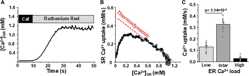
The type 2a sarcoplasmic/endoplasmic reticulum Ca2+-ATPase (SERCA2a) plays a central role in Ca2+ signaling of cardiomyocytes. The speed at which SERCA2a pumps Ca2+ from the cytosol into the sarcoplasmic reticulum (SR) determines the diastolic relaxation rate. SERCA2a activity also sets SR Ca2+ load, which determines the amplitude of SR Ca2+ release and the systolic contraction strength. While SERCA2a controls the SR luminal [Ca2+] ([Ca2+]SR), less is known about how dynamic changes in [Ca2+]SR affect SERCA2a function. By measuring the endoplasmic reticulum [Ca2+] ([Ca2+]ER) with the Ca2+ sensor R-CEPIA1er, we characterized the function of recombinant human and native mouse SERCA2a. We found that despite low endoplasmic reticulum (ER) Ca2+ gradient, SERCA2a-mediated Ca2+ transport was significantly slower at low [Ca2+]ER than at intermediate [Ca2+]ER. It appears that certain [Ca2+]ER is required for optimal SERCA2a Ca2+ transport. We tested whether negatively charged amino acids within the luminal loop between transmembrane helices M7 and M8 contribute to SERCA2a regulation by [Ca2+]ER. We found that the triple mutation E877L/D878L/E883L in the M7-M8 loop reduces SERCA2a Ca2+ transport particularly at intermediate [Ca2+]ER. Destabilizing the M7-M8 loop by breaking a disulfide bond between cysteines 875 and 887 abolished ER Ca2+ transport. Complementary molecular dynamics simulations showed that the triple mutant E877L/D878L/E883L stabilizes a Ca2+-bound E2 state of the pump, slowing down release of Ca2+ from the transport sites into the ER compared with the wild-type SERCA2a. These results revealed, for the first time, that SERCA2a Ca2+ transport is regulated by the luminal Ca2+ by interacting with the M7-M8 loop.

Keywords: calcium ATPase; calcium pump; endoplasmic reticulum; fluorescent microscopy; intracellular calcium signaling.


Figures

Fig. 1.
Fig. 1.
SERCA2a-mediated Ca2+ uptake as a function of [Ca2+]ER in HEK239 cells expressing human SERCA2a. A) Experimental protocol to measure SERCA-mediated ER Ca2+ uptake. A line-scan image of R-CEPIA1er (top) and corresponding profile of changes in [Ca2+]ER (bottom) were measured in control conditions, during RyR2 activation with 5 mM caffeine with following RyR2 inhibition with 15 μM ruthenium red. In the end of each experiment, the R-CEPIA1er signal was converted to [Ca2+] after measuring Fmax in the presence 5 mM Ca2+ and 5 μM ionomycin and Fmin in the presence of 5 mM caffeine. Black line (Ctrl)—control conditions. Red line (TG)—thapsigargin. B) For each individual cell, ER Ca2+ uptake was analyzed as the first derivative (d[Ca2+]ER/dt) and plotted as a function of ER Ca2+ load ([Ca2+]ER). C) The average results of SERCA2a-mediated Ca2+ uptake rate at different ER Ca2+ loads: low Ca2+ loads were within the range 0.05–0.10 mM; intermediate (inter) loads were within 0.2–0.4 mM and high loads were within 0.6–0.8 mM. The analysis is based on results from 40 cells.
Fig. 2.
Fig. 2.
SERCA-mediated ER Ca2+ uptake during Ca2+ waves activated by relieving RyR2 inhibition. A) An example of Ca2+ waves recorded after relieving RyR2 inhibition by 10 mM cytosolic [Mg2+]. B) The average results of SERCA2a-mediated Ca2+ uptake rate during Ca2+ wave at different ER Ca2+ loads: low 0.05–0.10 mM; intermedium (inter) 0.2–0.4 mM and high 0.5–0.6 mM. The analysis is based on results from 20 cells.
Fig. 3.
Fig. 3.
SERCA2a-mediated Ca2+ uptake as a function of [Ca2+]SR in mouse ventricular myocytes. A) Profile of changes in [Ca2+]SR during RyR2 activation with 5 mM caffeine with following RyR2 inhibition with 15 μM ruthenium red. The R-CEPIA1er signal was converted to [Ca2+] after measuring Fmax in the presence 5 mM Ca2+ and 5 μM ionomycin and Fmin in the presence of 5 mM caffeine. B) For each individual cell, SR Ca2+ uptake was analyzed as the first derivative (d[Ca2+]ER/dt) and plotted as a function of [Ca2+]ER. C) The average results of SERCA2a-mediated Ca2+ uptake rate at different SR Ca2+ loads: low 0.05–0.10 mM; intermedium (inter) 0.2–0.4 mM and high 0.6–0.8 mM. The analysis is based on results from 11 myocytes.
Fig. 4.
Fig. 4.
SERCA2a-mediated Ca2+ uptake as a function of [Ca2+]ER in HEK239 cells expressing SERCA2aWT and three M7-M8 SERCA2a mutants. A) Left: analysis of the electrostatic potential SERCA2a map. Negative charges are labeled in red. Positive charges are labeled in blue. Right: position of two cysteines (green) and five NCA (red) in the M7–M8 loop. The list of three SERCA2a mutants with NCAs and single cysteine mutations. B) Changes in [Ca2+]SR during RyR2 activation with following RyR2 inhibition in HE293 cells expressing SERCA2aWT and different SERCA2a mutants: SERCA2aC875A, SERCA2a5NCA, and SERCA2a3NCA. The R-CEPIA1er signal was converted to [Ca2+] after measuring Fmax in the presence 5 mM Ca2+ and 5 μM ionomycin and Fmin in the presence of 5 mM caffeine. C) ER Ca2+ uptake was analyzed as the first derivative (d[Ca2+]ER/dt) and plotted as a function of [Ca2+]ER. D) The average results of maximal ER Ca2+ uptake rate in HE293 cells expressing SERCA2aWT and different SERCA2a mutants. The analysis is based on results from 40 cells for SERCA2aWT, 9 cells for SERCA2a5NCA and 24 cells for SERCA2a3NCA.
Fig. 5.
Fig. 5.
Representation of translocation of a single Ca2+ ion from the transport sites of SERCA2a into the SR lumen. Structures of the SERCA2a embedded in a lipid bilayer extracted from the MD trajectories of the WT SERCA bound to a single Ca2+ ion. The head groups of the lipid bilayer are shown as white spheres; SERCA2a is shown as a gray cartoon representation. Key SERCA2a residues and the Ca2+ ion are shown as a van der Waals representation. We note that this translocation mechanism is inhibited upon mutation of residues E877, D878, and E883 to leucine.
Fig. 6.
Fig. 6.
Distance evolution between residues E90 and E309 with the Ca2+ ion occluded in the transport sites in the WT and triple mutant SERCA2a. Distances were calculated from each independent MD trajectory between the carbon delta (Cδ) of residues E90 (black trace) or E309 (red trace) and the Ca2+ ion. For these simulations, we used the crystal structure of SERCA2a representing the state at which Ca2+ is released into the SR lumen (PDB: 3b9b). This structure, which was obtained in the absence of Ca2+, a single Mg2+ ion was resolved in the transport sites; we replaced this ion for Ca2+ as a starting point for performing the atomistic MD simulations. The plots show that in SERCA2aWT, Ca2+ partially (e.g. replicates 1 and 3) or fully (e.g. replicate 2) translocate into the luminal side of the lipid bilayer. Translocation is inhibited upon mutation of residues E877, D878, and E883 to leucine.

References

    1. Bers DM. 2002. Cardiac excitation-contraction coupling. Nature. 415:198–205. - PubMed
    1. Kranias EG, Hajjar RJ. 2012. Modulation of cardiac contractility by the phospholamban/SERCA2a regulatome. Circ Res. 110:1646–1660. - PMC - PubMed
    1. Olesen C, et al. . 2007. The structural basis of calcium transport by the calcium pump. Nature. 450:1036–1042. - PubMed
    1. Bovo E, et al. . 2020. Dimerization of SERCA2a enhances transport rate and improves energetic efficiency in living cells. Biophys J. 119:1456–1465. - PMC - PubMed
    1. Currie S, Smith GL. 1999. Enhanced phosphorylation of phospholamban and downregulation of sarco/endoplasmic reticulum Ca2+ ATPase type 2 (SERCA 2) in cardiac sarcoplasmic reticulum from rabbits with heart failure. Cardiovasc Res. 41:135–146. - PubMed