. 2025 Dec;14(1):2465010.
doi: 10.1080/2162402X.2025.2465010. Epub 2025 Feb 17.

IL-15 transpresentation by ovarian cancer cells improves CD34+ progenitor-derived NK cell's anti-tumor functionality

Affiliations

IL-15 transpresentation by ovarian cancer cells improves CD34+ progenitor-derived NK cell's anti-tumor functionality

M Vidal-Manrique et al. Oncoimmunology. 2025 Dec.

Abstract

Ovarian cancer (OC) is the most lethal gynecological malignancy. As high numbers of Natural Killer (NK) cells in ascites associate with improved survival, the adoptive transfer of allogeneic NK cells is an attractive therapeutic strategy. An approach to further improve NK cell expansion and anti-tumor functionality post-infusion includes IL-15 transpresentation (transIL-15), which involves surface expression of the IL-15 cytokine bound to IL-15Rα. However, others have substantiated that systemic administration of ALT/N-803, a soluble molecule mimicking transIL-15, leads to T cell-mediated rejection of the infused allogeneic NK cell product. In addition, whether transIL-15 induce superior expansion and functionality of our hematopoietic progenitor cell-derived NK cells (HPC-NK) remains understudied. Here, we propose to transfect OC cells with IL-15 and IL-15Rα mRNA and evaluate HPC-NK cell stimulation in vitro. Co-transfection of both mRNAs resulted in surface co-expression of both components, thus mimicking the transIL-15. Importantly, co-culture of HPC-NK cells with transIL-15 OC cells resulted in superior proliferation, IFNγ production, cytotoxicity and granzyme B secretion. Furthermore, we observed uptake of IL-15Rα by HPC-NK cells when co-cultured with transIL-15 OC cells, which associates with NK cell long-term proliferation and survival. Superior killing and granzyme B secretion were also observed in transIL-15 OC spheroids. Our results demonstrate that local delivery of IL-15 and IL-15Rα mRNA to OC tumors may be a safer strategy to boost HPC-NK cell therapy of OC through IL-15 transpresentation.

Keywords: IL-15 transpresentation; NK cell immunotherapy; mRNA delivery; ovarian cancer.


Conflict of interest statement

Roland Brock is co-founder and co-owner of Mercurna B. V. and RIBOPRO B. V., companies that develop mRNA-based therapeutics.

Figures
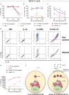
Figure 1.
Figure 1.
IL-15 and IL-15Rα mRNA are effectively transfected in SKOV-3. Comparison of (a) viability, (b) IL-15Rα+ percentage and (c) IL-15Rα+ MFI of transfected cells measured at the indicated time points. (d) Representative dot plots showing co-expression of IL-15 and IL-15Rα 24 h after transfection with non-washed (upper row) and washed (bottom row) mRNAs. Data derives from 3 independent replicates. (e) IL-15 secretion data from 2–6 independent replicates from one experiment. For all graphs, lipofectamine nanoparticles were either washed 2 h after transfection (dotted lines) or not (continuous lines). (f) Model depicting secretion mechanism of IL-15 by IL-15-transfected OC cells (left) or transIL-15 OC cells (right). All data represents mean values ± SEM. Statistics derive from two-way ANOVA with Bonferroni correction comparing washed and non-washed samples within the same transfection condition and same time point (*p < 0.05, ****p < 0.0001).
Figure 2.
Figure 2.
HPC-NK cells show superior functionality upon co-culture with transIL-15-transfected SKOV-3 cells. (a) Percentage of the indicated markers on 8 independent HPC-NK cell donors. (b) Fold change proliferation of HPC-NK cells 3 days after incubation with transfected SKOV-3 cells compared to unstimulated HPC-NK cells alone, and representative histograms showing loss of eFluor450. Data comes from 4 independent experiments, 4 independent fresh donors. (c) Percentage of CD107a+, IFNγ+ and TNF+ in viable CD56+ HPC-NK cultured for 4 h with targets whose media either contains secreted IL-15 or (d) was refreshed just prior to HPC-NK addition. Data comes from 3 independent experiments, 6 independent donors. (e) Representative flow cytometry dot plots of CD107a (y-axis) and IFNγ (x-axis) in HPC-NK cells cultured with transfected SKOV-3 cells whose media was removed (refreshed medium), or not (IL-15-containing medium), prior to plating of the effectors. Numbers represent percentage of positive cells per quadrant. (f) Percentage of target killing and (g) IFNγ and (h) granzyme B secreted by HPC-NK cells after 16-20 h co-culture. Results derive from 2 independent experiments, 4 independent HPC-NK donors. Bars represent mean values ± SEM. Whenever applicable, one or two-way ANOVA within each potency readout or effector-to-target ratio were used for statistical analysis with Bonferroni correction (*p < 0.05, **p < 0.01, ***p < 0.001, ****p < 0.0001).
Figure 3.
Figure 3.
High grade serous OVCAR-4 cells and primary-derived OC cells are effectively transfected and activate HPC-NK cell proliferation and potency. (a) Viability, (b) percentage of IL-15Rα+ cells and MFI within IL-15Rα+ cells and (c) IL-15 secretion of transfected and washed OVCAR-4 cells at the indicated time points. (d) Representative flow cytometry histograms showing loss of eFluor450 after three days in one fresh HPC-NK cell donor. (e) Percentage of CD107a+, IFNγ+ and TNF+ in viable CD56+ HPC-NK cells after 4 h co-culture with OVCAR-4 cells. Data derives from 4 independent donors, one experiment. (f) Viability, (g) percentage of IL-15Rα+ cells and MFI within IL-15Rα+ cells, and (h) IL-15 secretion of transfected and washed ASC009 cells at the indicated time points. (i) Representative flow cytometry histograms showing loss of eFluor450 after three days in one fresh HPC-NK cell donor. (j) Percentage of CD107a+, IFNγ+ and TNF+ in viable CD56+ HPC-NK cells after 4 h co-culture with ASC009 cells. All viability and transfection data comes from 3 independent replicates, one experiment. All potency data derives from 4 independent HPC-NK donors, one experiment. Data represent mean values ± SEM. Two-way ANOVA with Bonferroni correction comparing all conditions within the same time point or potency readout was used (*p < 0.05, **p < 0.01, ***p < 0.001, ****p < 0.0001).
Figure 4.
Figure 4.
SKOV-3 OC spheroids are effectively transfected with transIL-15, and results in superior HPC-NK-mediated cytotoxicity. (a) Representative image of gfp-transfected, non-washed SKOV-3 spheroids 24 h after transfection (scale bar represents 100 μm). (b) Viability, (c) percentage of IL-15Rα+, (d) MFI of IL-15Rα+ and (e) IL-15 secretion of SKOV-3 spheroids 24 h after transfection. Data comes from 6 independent spheroids, 4 independent experiments. (f) Percentage of target cell death and (g) granzyme B secretion by HPC-NK cells co-cultured with spheroids for 16-20 h. For each donor, data comes from 2 independent experiments, 4 independent replicates per experiment. All data represent mean values ±SD. When applicable, student t-tests or two-way ANOVA with Bonferroni correction to compare different transfection conditions within the same effector-to-target ratios were used (*p < 0.05, **p < 0.01, ***p < 0.001, ****p < 0.0001).

References

    1. Momenimovahed Z, Tiznobaik A, Taheri S, Salehiniya H.. ovarian cancer in the world: epidemiology and risk factors. Int J Womens Health. 2019;11:287–11. doi:10.2147/IJWH.S197604. - DOI - PMC - PubMed
    1. Timmermans M, Sonke GS, Van de Vijver KK, van der Aa MA, Kruitwagen RFPM. No improvement in long-term survival for epithelial ovarian cancer patients: a population-based study between 1989 and 2014 in the Netherlands. Eur J Cancer. 2018;88:31–37. doi:10.1016/j.ejca.2017.10.030. - DOI - PubMed
    1. Laskowski TJ, Biederstadt A, Rezvani K. Natural killer cells in antitumour adoptive cell immunotherapy. Nat Rev Cancer. 2022;22(10):557–575. doi:10.1038/s41568-022-00491-0. - DOI - PMC - PubMed
    1. Hoogstad-van Evert JS, Maas RJ, van der Meer J, Cany J, van der Steen S, Jansen JH, Miller JS, Bekkers R, Hobo W, Massuger L, et al. Peritoneal NK cells are responsive to IL-15 and percentages are correlated with outcome in advanced ovarian cancer patients. Oncotarget. 2018;9(78):34810–34820. doi:10.18632/oncotarget.26199. - DOI - PMC - PubMed
    1. de Jonge P, van Hauten PMM, Janssen LD, de Goede AL, Berrien-Elliott MM, van der Meer JMR, Mousset CM, Roeven MWH, Foster M, Blijlevens N, et al. Good manufacturing practice production of CD34(+) progenitor-derived NK cells for adoptive immunotherapy in acute myeloid leukemia. Cancer Immunol Immunother. 2023;72(10):3323–3335. doi:10.1007/s00262-023-03492-6. - DOI - PMC - PubMed

MeSH terms

LinkOut - more resources