. 2025 Feb 17;16(1):1703.
doi: 10.1038/s41467-025-56876-w.

RNA methyltransferase SPOUT1/CENP-32 links mitotic spindle organization with the neurodevelopmental disorder SpADMiSS

Avinash V Dharmadhikari #  1   2 Maria Alba Abad #  3 Sheraz Khan #  4   5   6   7 Reza Maroofian  8 Tristan T Sands  9 Farid Ullah  4   5 Itaru Samejima  3 Yanwen Shen  10   11   12   13 Martin A Wear  14 Kiara E Moore  4   5 Elena Kondakova  15 Natalia Mitina  15 Theres Schaub  16 Grace K Lee  17 Christine H Umandap  18   19 Sara M Berger  19 Alejandro D Iglesias  19 Bernt Popp  20 Rami Abou Jamra  20 Heinz Gabriel  21 Stefan Rentas  22 Alyssa L Rippert  23 Christopher Gray  23 Kosuke Izumi  23 Laura K Conlin  24 Daniel C Koboldt  25   26 Theresa Mihalic Mosher  27 Scott E Hickey  26   28 Dara V F Albert  26   29 Haley Norwood  30 Amy Feldman Lewanda  31 Hongzheng Dai  32   33 Pengfei Liu  32   33 Tadahiro Mitani  32 Dana Marafi  32   34 Hatice Koçak Eker  35 Davut Pehlivan  32   36   37 Jennifer E Posey  32 Natalie C Lippa  38 Natalie Vena  38 Erin L Heinzen  39   40 David B Goldstein  41 Cyril Mignot  42 Jean-Madeleine de Sainte Agathe  43 Nouriya Abbas Al-Sannaa  44 Mina Zamani  45   46 Saeid Sadeghian  47 Reza Azizimalamiri  47 Tahere Seifia  45   46 Maha S Zaki  48 Ghada M H Abdel-Salam  48 Mohamed S Abdel-Hamid  49 Lama Alabdi  50 Fowzan Sami Alkuraya  50 Heba Dawoud  51 Aya Lofty  51 Peter Bauer  52 Giovanni Zifarelli  52 Erum Afzal  53 Faisal Zafar  53 Stephanie Efthymiou  8 Daniel Gossett  54   55 Meghan C Towne  27 Raey Yeneabat  56 Belen Perez-Duenas  57   58   59 Ana Cazurro-Gutierrez  58   59 Edgard Verdura  58   60 Veronica Cantarin-Extremera  61   62 Ana do Vale Marques  63 Aleksandra Helwak  3 David Tollervey  3 Sandeep N Wontakal  56 Vimla S Aggarwal  64 Jill A Rosenfeld  32 Victor Tarabykin  15   16 Shinya Ohta  65 James R Lupski  32   36   66 Henry Houlden  8 William C Earnshaw  3 Erica E Davis  67   68 A Arockia Jeyaprakash  69   70 Jun Liao  71
Affiliations

RNA methyltransferase SPOUT1/CENP-32 links mitotic spindle organization with the neurodevelopmental disorder SpADMiSS

Avinash V Dharmadhikari et al. Nat Commun. .

Abstract

SPOUT1/CENP-32 encodes a putative SPOUT RNA methyltransferase previously identified as a mitotic chromosome associated protein. SPOUT1/CENP-32 depletion leads to centrosome detachment from the spindle poles and chromosome misalignment. Aided by gene matching platforms, here we identify 28 individuals with neurodevelopmental delays from 21 families with bi-allelic variants in SPOUT1/CENP-32 detected by exome/genome sequencing. Zebrafish spout1/cenp-32 mutants show reduction in larval head size with concomitant apoptosis likely associated with altered cell cycle progression. In vivo complementation assays in zebrafish indicate that SPOUT1/CENP-32 missense variants identified in humans are pathogenic. Crystal structure analysis of SPOUT1/CENP-32 reveals that most disease-associated missense variants are located within the catalytic domain. Additionally, SPOUT1/CENP-32 recurrent missense variants show reduced methyltransferase activity in vitro and compromised centrosome tethering to the spindle poles in human cells. Thus, SPOUT1/CENP-32 pathogenic variants cause an autosomal recessive neurodevelopmental disorder: SpADMiSS (SPOUT1 Associated Development delay Microcephaly Seizures Short stature) underpinned by mitotic spindle organization defects and consequent chromosome segregation errors.


Conflict of interest statement

Competing interests: BCM and Miraca Holdings have formed a joint venture with shared ownership and governance of Baylor Genetics (BG), which performs clinical microarray analysis (CMA), clinical ES (cES), and clinical biochemical studies. JRL serves on the Scientific Advisory Board of the BG. The Department of Molecular and Human Genetics at Baylor College of Medicine receives revenue from clinical genetic testing conducted at BG Laboratories. JRL has stock ownership in 23andMe, is a paid consultant for Genomics International, and is a coinventor on multiple United States and European patents related to molecular diagnostics for inherited neuropathies, eye diseases, genomic disorders, and bacterial genomic fingerprinting. DP provides consulting service for Ionis Pharmaceuticals. The other authors declare no competing interests.

Figures
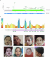
Fig. 1
Fig. 1. Majority of SPOUT1/CENP-32 variants identified in affected individuals are located in the RNA methyltransferase domain of the protein and at amino acid residues intolerant to variation.
A Schematic of SPOUT1/CENP-32 at the exon level (NM_016390.4) and protein level (NP_057474.2) with positions of the variants identified in our cohort and the number of alleles in unrelated families identified for each variant. Missense variants are displayed in green, loss-function variants in black and the in-frame deletion in red. The single annotated domain in green is from Pfam and reflects the RNA methyltransferase domain. B Overlay of SPOUT1/CENP-32 variants identified in this cohort with the protein intolerance landscape of SPOUT1/CENP-32 from MetaDome. MetaDome analyses shows the mutation tolerance at each position of the protein, with red being intolerant, yellow is neutral and blue is tolerant. CJ Variable dysmorphic features were seen in 50% (14/28) individuals including high arched palate, prominent ears, upturned nostrils, tented upper lip and high forehead. C Subject: family G-II-1 at 1 year. D Subject: family G-II-1 at 9 years. E Subject: family G-II-2 at 6 years. F Subject: family L-II-1 at 8 years. G Subject: family L-II-2 at 1 year 4 months. H Subject: family M-V-2 at 4 years. I Subject: family N-IV-2 at 18 months. J Subject: family Q-II-1 at 9 years. Created in BioRender.
Fig. 2
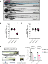
Fig. 2. spout1/cenp-32 mutant larvae display reduced head area.
A Representative bright field lateral images of spout1/cenp-32+/+, spout1/cenp-32+/- and spout1/cenp-32−/− are shown at 3 days post-fertilization (dpf); pink dashed outline depicts head size measured, and the blue dotted line shows the body length measured. B, C Quantification of lateral head size (B) and body length (C) measurements were analyzed from combined experimental batches (n = 2 biological replicates). Statistical differences were calculated using a non-parametric ANOVA with Kruskal-Wallis test followed by Dunn’s multiple comparisons test by controlling False Discovery Rate (original FDR method of Benjamini and Hochberg); ( + ) and (-) indicate significant and non-significant differences, respectively. Median values are shown with pink horizontal lines. See Supplementary Table S2 for exact adjusted q-values and numbers of larvae. D Schematic representation (left) of sample preparation for qPCR to monitor endogenous spout1/cenp-32 transcript (right); statistical differences were calculated using unpaired t-test. Two biological replicates were performed, each with technical triplicates. Scale bar, 300 µM. Source data are provided as a Source Data file.
Fig. 3
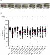
Fig. 3. In vivo complementation assay indicates that variants identified in affected individuals are pathogenic.
A Representative bright field images of uninjected control (UC), morphants (MO), and MO plus mRNA-injected larvae at 3 dpf. B In vivo complementation data show that variants identified in affected individuals are pathogenic. p.T130R, rs6478854 is a presumed benign variant used as a negative control. Scale bar, 300 µM. Statistical differences were calculated using non-parametric ANOVA with Kruskal-Wallis test followed by Dunn’s multiple comparisons test by controlling False Discovery Rate (original FDR method of Benjamini and Hochberg); ( + ) and (-) indicate significant and non-significant differences, respectively. Median values are shown with pink horizontal lines. See Supplementary Table S2 for exact adjusted q-values and numbers of larvae. Source data are provided as a Source Data file.
Fig. 4
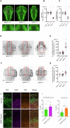
Fig. 4. spout1/cenp-32-/- mutant larvae have CNS defects, apoptosis and altered cell cycle progression.
A Representative dorsal images of wholemount larvae fixed at 3 dpf and immunostained for acetylated tubulin to demarcate axon tracts. We counted commissural axons crossing the midline between the optic tecta (pink dashed line); and the area of optic tecta (pink dashed oval). Dashed white box (upper panel) represents magnified image in lower panel. Scale bar, 100 µM. B, C Quantification of optic tecta size and intertectal neuron number, respectively. D Representative dorsal inverted fluorescent images of zebrafish larvae marked by TUNEL at 2 dpf. The region of interest (ROI) quantified is shown with a red rectangle. Scale bar, 100 µM. E Quantification of TUNEL stained cells as measured in the ROI. F Representative dorsal inverted fluorescent images showing phospho-histone H3 (pHH3) positive cells at 2 dpf. The ROI quantified is shown with a red rectangle. Scale bar, 100 µM. G Quantification of pHH3 positive cells as measured in ROI. Data shown in B, C, E, and G are combined from two biological replicates. Statistical differences were calculated using non-parametric ANOVA with Kruskal-Wallis test followed by Dunn’s multiple comparisons test by controlling False Discovery Rate (original FDR method of Benjamini and Hochberg); ( + ) and (-) indicate significant and non-significant differences, respectively. Median values are shown with pink horizontal lines. See Supplementary Table S2 for exact adjusted q-values and numbers of larvae. H, I Murine cortical progenitor cells electroporated with shRNA against Spout1 less frequently leave mitotic cell cycle both within 24 hours (I, left diagram), and 48 hours (I, right diagram) after electroporation, and also less frequently migrate to the cortical plate (CP) (white arrows) compared to wild type cells (Note: GFP positive neurons in the control CP and lack of them in shRNA electroporated brains). In utero electroporation of targeting constructs into the lateral ventricles was carried out at E13.5. 24 hours later, neocortical progenitors were labeled with BrdU during S phase of the mitotic cycle. Neocortical cells that express both mitotic marker Ki67 and GFP represent a fraction of the cortical cells that still proliferate 48 hours after in utero electroporation, while cells that express BrdU, Ki67 and GFP represent a fraction of cells that proliferate 24 hours after BrdU injection. Statistical differences were calculated using two-sided unpaired t-test (*, P ≤ 0.05; ****, P ≤ 0.0001). I left diagram P = 0.0176. Data are presented as Mean +/- SD. Number of brain samples analyzed: 3 (control), 4 (experiment); number of slices analyzed: 9 (control), 13 (experiment). Scale bar, 50 µM. Source data are provided as a Source Data file. Created in BioRender.
Fig. 5
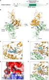
Fig. 5. Structural characterization reveals that SPOUT1/CENP-32 is a SPOUT Methyltransferase that dimerizes through its catalytic domain.
A Domain architecture of SPOUT1/CENP-32 with the structural domains of SPOUT1/CENP-32 highlighted. B Cartoon representation of the crystal structure of SPOUT1/CENP-32 71-376 bound to S-adenosyl homocysteine (SAH) in two orientations (left and right panel). The main domains of SPOUT1/CENP-32 (SPOUT domain and OB-fold) are highlighted. SAH is bound to the cofactor pocket, indicating that SPOUT1/CENP-32 could be an active methyltransferase. Our structural analysis is consistent with the structure that was deposited by SGC while this manuscript was in preparation (PDB: 4RG1). C, D and F Close-up of the active site of SPOUT1/CENP-32 with SAH (C and D) and with SAM (F) showing the electron density map for SAH (C) and the amino acid residues that are responsible for the interactions that stabilize cofactor binding. The binding of SAH and SAM is similar. E Close-up of the electrostatic surface potential of SPOUT1/CENP-32 71-376 bound to SAM showing the deep pocket formed by the trefoil knot.
Fig. 6
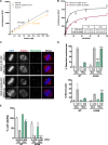
Fig. 6. SPOUT1/CENP-32 is an active Methyltransferase and its activity is crucial for its mitotic function.
A Titration of methyltransferases (SPOUT1/CENP-32 and NSUN6) to assess the methyltransferase activity of SPOUT1/CENP-32 in vitro where Survivin protein was used as a negative control. Increasing amounts of either methyltransferase (NSUN6, a well-known RNA methyltransferase, or SPOUT1/CENP-32) or Survivin protein (0–20 μM) were incubated with 1 μg of total RNA extract (isolated from NSUN6 depleted or SPOUT1/CENP-32 depleted U2OS cells) and 20 μM SAM for 30 min at room temperature. RLU (Relative Luminescence unit). Data from three biological replicates, n = 9. Data represented as mean ± SEM. Source data are provided as a Source Data file. B Methyltransferase assay to determine the Km and Vmax of SPOUT1/CENP-32 WT and A356N in the presence of constant SAM (20 μM) and increasing amounts of a GAPDH mRNA hairpin as substrate (GCCCCCUCUGCUGAUGCCCCCAUGUUCGUCAUGGGUGUGAA; 0 – 100 μM). Km and Vmax values for SPOUT1/CENP-32 proteins are depicted in the table. RLU (Relative Luminescence unit). Data from three biological replicates, n = 6. Data represented as mean ± SEM. Source data are provided as a Source Data file. C Representative immunofluorescence images of SPOUT1/CENP-32 inducible U2OS cell lines for the analysis of centrosome detachment upon Control (siRNA C) or SPOUT1/CENP-32 (siRNA C32) depletion using siRNA oligos and rescue with either induction of SPOUT1/CENP32 WT-GFP or SPOUT1/CENP32 A356N-GFP expression. Conditions with doxycycline only. Conditions without doxycycline in Fig. S15F. D Quantification for the analysis of the centrosome detachment phenotype (% of cells with detached centrosomes; top panel) and chromosome segregation errors (% of micronuclei; bottom panel) in inducible U2OS cell lines expressing SPOUT1/CENP-32 WT-GFP or SPOUT1/CENP-32 A356N-GFP. Data are representative of a minimum of three biological replicates, mean ± SD, n = 3. Scale bar, 10 μm; Two-sided Fisher’s exact test with Bonferroni correction (****, P ≤ 0.0001). Exact p-values for the detached centrosome phenotype vs the WT condition are: A356N siRNA C +Dox=2.42E-06, A356N siRNA C32 +Dox=7.90E-20. Exact p-values for the micronuclei phenotype vs the WT condition are: A356N siRNA C32 +Dox=1.43E-11. Data points for each of the replicates are shown. Source data are provided as a Source Data file. E Cell viability evaluation of inducible U2OS cell lines expressing SPOUT1/CENP-32 WT-GFP or SPOUT1/CENP-32 A356N-GFP assessed by the MTT assay. Three independent biological replicates, mean ± SEM. WT siRNA C -Dox: n = 26; WT siRNA C32 -Dox; n = 26; WT siRNA C +Dox: n = 25; WT siRNA C32 +Dox: n = 27; A356N siRNA C -Dox: n = 11; A356N siRNA C32 -Dox: n = 18; A356N siRNA C +Dox: n = 13; A356N siRNA C32 +Dox: n = 18. Non-parametric ANOVA with Kruskal-Wallis followed by Dunn’s multiple comparisons test (***, P ≤ 0.001; ****, P ≤ 0.0001). Exact p-values vs the WT siRNA C -Dox condition are: WT siRNA C32 -Dox=1.44E-04, A356N siRNA C32 -Dox=1E-06, A356N siRNA C32 +Dox=1E-06. Source data are provided as a Source Data file.
Fig. 7
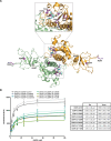
Fig. 7. SPOUT1/CENP-32 patient variants show decreased methyltransferase activity.
A Mapping of the SPOUT1/CENP-32 variants in the SPOUT1/CENP-32 71-376 SAH-bound structure. Variant residues are highlighted in purple. B Methyltransferase assay to determine the Km and Vmax of SPOUT1/CENP-32 WT and the variants in the presence of constant SAM (20 μM) and increasing amounts of a GAPDH mRNA hairpin as substrate (GCCCCCUCUGCUGAUGCCCCCAUGUUCGUCAUGGGUGUGAA; 0 – 100 μM; left panel). Km and Vmax values for all SPOUT1/CENP-32 proteins are depicted in the table (right panel). R square values are as follows: SPOUT1/CENP-32 WT – 0.79 (n = 10), SPOUT1/CENP-32 N86D – 0.83 (n = 6), SPOUT1/CENP32 G98S – 0.87 (n = 6), SPOUT1/CENP-32 G244S – 0.93 (n = 10), SPOUT1/CENP-32 T289M – 0.97 (n = 10), SPOUT1/CENP-32 G293S – 0.93 (n = 10), SPOUT1/CENP-32 T353M – 0.9 (n = 10), SPOUT1/CENP-32 T130R – 0.98 (n = 4). Data from three independent biological replicates, mean ± SD. Source data are provided as a Source Data file.
Fig. 8
Fig. 8. SPOUT1/CENP-32 patient variants lead to alterations of spindle organisation.
A Representative fluorescence images of a Control (siRNA C) or SPOUT1/CENP-32 (siRNA C32) siRNA rescue assay in stable U2OS cell lines with inducible expression of different SPOUT1/CENP-32-GFP constructs. Conditions with doxycycline only; conditions without doxycycline in Fig. S17A. B, C Quantification for the analysis of the centrosome detachment phenotype (% of cells with detached centrosomes; B) and chromosome segregation errors (% of micronuclei; C) in inducible U2OS cell lines expressing SPOUT1/CENP-32 WT-GFP or the SPOUT1/CENP-32 patient variants. For easy direct comparison with the SPOUT1/CENP-32 variant conditions, the SPOUT1/CENP-32 WT conditions shown in Fig. 6D are also shown in this figure. Data are representative of a minimum of three biological replicates, n ≥ 70 cells analyzed in total per treatment, mean ± SD, n = 3. Scale bar, 10 μm. Two-sided Fisher’s exact test with Bonferroni correction (*, P ≤ 0.05; **, P ≤ 0.01; ***, P ≤ 0.001; ****, P ≤ 0.0001). Exact p-values for the detachment centrosome phenotype (B) vs WT condition are: N86D siRNA C +Dox=3.1E-03, N86D siRNA C32 +Dox=4.3E-30, G98S siRNA C +Dox=1.9E-02, G98S siRNA C32 +Dox=4.9E-33, G244S siRNA C32 +Dox=4.8E-15, T289M siRNA C32 +Dox=1.8E-25, G293S siRNA C32 +Dox=2.7E-30, T353M siRNA C +Dox=3.3E-07, T353M siRNA C32 +Dox=4.1E-28. Exact p-values for the micronuclei phenotype (C) vs WT condition are: N86D siRNA C +Dox=1.3E-02, N86D siRNA C32 +Dox=2.9E-04, G98S siRNA C32 -Dox=1.9E-07, G98S siRNA C +Dox=2.5E-04, G98S siRNA C32 +Dox=2.1E-05, G244S siRNA C32 +Dox=1.6E-15, T289M siRNA C32 +Dox=1.2E-24, G293S siRNA C32 +Dox=1.3E-03, T353M siRNA C +Dox=1.1E-04, T353M siRNA C32 +Dox=5.5E-37. Data points for each of the replicates are shown. Source data are provided as a Source Data file. D Cell viability evaluation of inducible U2OS cell lines expressing SPOUT1/CENP-32 WT-GFP or SPOUT1/CENP-32 patient variants assessed by the MTT assay. For easy direct comparison with the SPOUT1/CENP-32 variant conditions, the SPOUT1/CENP-32 WT conditions shown in Fig. 6E are also shown in this figure. Three independent biological replicates, mean ± SEM. WT siRNA C -Dox: n = 26; WT siRNA C32 -Dox; n = 26; WT siRNA C +Dox: n = 25; WT siRNA C32 +Dox: n = 27; N86D siRNA C -Dox: n = 22; N86D siRNA C32 -Dox: n = 25; N86D siRNA C +Dox: n = 27; N86D siRNA C32 +Dox: n = 22; G98S siRNA C -Dox: n = 27; G98S siRNA C32 -Dox; n = 26; G98S siRNA C +Dox: n = 27; G98S siRNA C32 +Dox: n = 24; G244S siRNA C -Dox: n = 25; G244S siRNA C32 -Dox; n = 27; G244S siRNA C +Dox: n = 27; G244S siRNA C32 +Dox: n = 27; T289M siRNA C -Dox: n = 18; T289M siRNA C32 -Dox; n = 18; T289M siRNA C +Dox: n = 18; T289M siRNA C32 +Dox: n = 18; T353M siRNA C -Dox: n = 18; T353M siRNA C32 -Dox; n = 18; T353M siRNA C +Dox: n = 18; T353M siRNA C32 +Dox: n = 18; G293S siRNA C -Dox: n = 27; G293S siRNA C32 -Dox; n = 27; G293S siRNA C +Dox: n = 27; G293S siRNA C32 +Dox: n = 27. Non-parametric ANOVA with Kruskal-Wallis followed by Dunn’s multiple comparisons test (*, p ≤ 0.05; **, p ≤ 0.01; ***, p ≤ 0.001; ****, p ≤ 0.0001). Exact p-values vs WT siRNA C -Dox condition are: WT siRNA C32 -Dox=1.4E-04, N86D siRNA C32 -Dox=2.4E-06, N86D siRNA C32 +Dox=4.6E-06, G98S siRNA C32 -Dox=8.7E-05, G98S siRNA C32 +Dox=2E-07, G244S siRNA C32 -Dox=1E-07, G244S siRNA C +Dox= 6.8E-03, G244S siRNA C32 +Dox=1E-07, T289M siRNA C32 -Dox=1E-07, T289M siRNA C32 +Dox= 1E-07, G293S siRNA C32 -Dox=1E-07, G293S siRNA C32 +Dox=1E-07, T353M siRNA C32 -Dox= 1E-07, T353M siRNA C +Dox=1.4E-02, T353M siRNA C32 +Dox=1E-07. Source data are provided as a Source Data file.
Fig. 9
Fig. 9. SPOUT1/CENP-32 bi-allelic variants disrupt centrosome-spindle pole tethering, causing chromosome segregation errors, altering cell cycle progress, and increasing apoptosis in neuronal progenitor cells.
Schematic summarizing the proposed differences between healthy cells and SPOUT1/CENP-32 variant cells leading to aneuploid daughter cells, altered cell cycle/ cell death, and microcephaly in affected individuals and mutant zebrafish.

Update of

  • RNA methyltransferase SPOUT1/CENP-32 links mitotic spindle organization with the neurodevelopmental disorder SpADMiSS.
    Dharmadhikari AV, Abad MA, Khan S, Maroofian R, Sands TT, Ullah F, Samejima I, Wear MA, Moore KE, Kondakova E, Mitina N, Schaub T, Lee GK, Umandap CH, Berger SM, Iglesias AD, Popp B, Jamra RA, Gabriel H, Rentas S, Rippert AL, Izumi K, Conlin LK, Koboldt DC, Mosher TM, Hickey SE, Albert DVF, Norwood H, Lewanda AF, Dai H, Liu P, Mitani T, Marafi D, Pehlivan D, Posey JE, Lippa N, Vena N, Heinzen EL, Goldstein DB, Mignot C, de Sainte Agathe JM, Al-Sannaa NA, Zamani M, Sadeghian S, Azizimalamiri R, Seifia T, Zaki MS, Abdel-Salam GMH, Abdel-Hamid M, Alabdi L, Alkuraya FS, Dawoud H, Lofty A, Bauer P, Zifarelli G, Afzal E, Zafar F, Efthymiou S, Gossett D, Towne MC, Yeneabat R, Wontakal SN, Aggarwal VS, Rosenfeld JA, Tarabykin V, Ohta S, Lupski JR, Houlden H, Earnshaw WC, Davis EE, Jeyaprakash AA, Liao J. Dharmadhikari AV, et al. medRxiv [Preprint]. 2024 Jan 9:2024.01.09.23300329. doi: 10.1101/2024.01.09.23300329. medRxiv. 2024. Update in: Nat Commun. 2025 Feb 17;16(1):1703. doi: 10.1038/s41467-025-56876-w. PMID: 38260255 Free PMC article. Updated. Preprint.

References

    1. Conduit, P. T., Wainman, A. & Raff, J. W. Centrosome function and assembly in animal cells. Nat. Rev. Mol. Cell Biol.16, 611–624 (2015). - PubMed
    1. Bornens, M. The centrosome in cells and organisms. Science335, 422–426 (2012). - PubMed
    1. Arquint, C., Gabryjonczyk, A. M. & Nigg, E. A. Centrosomes as signalling centres. Philos. Trans. R. Soc. Lond. B Biol. Sci.369, 20130464 (2014). - PMC - PubMed
    1. Heald, R., Tournebize, R., Habermann, A., Karsenti, E. & Hyman, A. Spindle assembly in Xenopus egg extracts: respective roles of centrosomes and microtubule self-organization. J. Cell Biol.138, 615–628 (1997). - PMC - PubMed
    1. Sir, J. H. et al. Loss of centrioles causes chromosomal instability in vertebrate somatic cells. J. Cell Biol.203, 747–756 (2013). - PMC - PubMed

MeSH terms