. 2025 Feb 17;16(1):1361.
doi: 10.1038/s41467-024-54949-w.

Time-resolved X-ray solution scattering unveils the events leading to hemoglobin heme capture by staphylococcal IsdB

Affiliations

Time-resolved X-ray solution scattering unveils the events leading to hemoglobin heme capture by staphylococcal IsdB

Omar De Bei et al. Nat Commun. .

Abstract

Infections caused by Staphylococcus aureus depend on its ability to acquire nutrients. One essential nutrient is iron, which is obtained from the heme of the human host hemoglobin (Hb) through a protein machinery called Iron-regulated surface determinant (Isd) system. IsdB is the protein in charge of heme extraction from Hb, which is the first step of the chain of events leading to iron transfer to the bacterium cell interior. In order to elucidate the molecular events leading from the formation of the initial IsdB:Hb complex to heme extraction, we use time-resolved X-ray solution scattering (TR-XSS) in combination with rapid mixing triggering. We succeed in defining the stoichiometry of IsdB:Hb binding and in describing the kinetics of the subsequent structural changes. The presented approach is potentially applicable to unveil the complex kinetic pathways generated by protein-protein interaction in different biological systems.


Conflict of interest statement

Competing interests: The authors declare no competing interests.

Figures

Fig. 1
Fig. 1. Schematic representation of the interaction between Staphylococcus aureus and human host for the bacterial acquisition of iron.
The bacterium produces hemolysins to lyse red blood cells (RBC) and release Hb. To proceed with the iron acquisition, S. aureus exposes the Hb receptor IsdB outside the cell wall. The magnified region shows how IsdB mediates the first step in iron acquisition: IsdB binds free Hb in the bloodstream, extracts heme and then passes it to the following proteins of the Isd system for its degradation and iron release.
Fig. 2
Fig. 2. SAXS patterns of IsdB:metHb and IsdB:oxyHb complexes measured at different protein concentrations.
a IsdB:metHb data (colored dots) together with fitted patterns (black solid lines) obtained with OLIGOMER. Legend reports concentrations in terms of monomeric complex (i.e. one IsdB molecule bound to a single Hb chain). b Schematic representation of the IsdB:metHb complex oligomeric equilibrium between IsdB molecules and metHb dimers (reagents) and two IsdB molecules bound to a metHb dimers (products, 2IsdB:metHbdim). c Volume fraction of the 2IsdB:metHbdim extracted from the SAXS data (closed symbols, error bars defined as s.e.m. (n = 10) are smaller than the symbol size) plotted as a function of total IsdB:metHb concentration (monomeric complex). The fit (continuous line) yields a dissociation constant KD = 1.68 ± 0.62 μM (see “Methods” and Supplementary Text). d IsdB:oxyHb data (colored dots) together with fitted patterns (black solid lines) obtained with OLIGOMER. Legend reports concentrations in terms of monomeric complex (i.e. one IsdB molecule bound to an Hb dimer). e Schematic representation of the IsdB:oxyHb complex oligomeric equilibrium in which oxyHb dimers with a single IsdB molecule bound to their β-chain (reagent, 1IsdB:βoxyHbdim) associate into a single particle (product, 2IsdB:βoxyHbtet). f Volume fraction of the 2IsdB:βoxyHbtet extracted from the SAXS data (closed symbols, error bars defined as s.e.m. (n = 10) are smaller than the symbol size) plotted as a function of total IsdB:oxyHb concentration (monomeric complex) together with the predicted dependence in the case of simple Hb dimer-to-tetramer association using the reported dissociation constant KD = 0.25 μM for oxyHb (continuous line) and the fit of the data (dashed line), which yields a dissociation constant of KD = 0.38 ± 1.30 μM.
Fig. 3
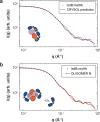
Fig. 3. Comparison between SAXS/WAXS merged data and calculated patterns based on molecular models.
a Data relative to the equilibrium complex formed by IsdB and metHb (closed symbols) vs. the 2IsdB:metHbdim calculated pattern obtained with CRYSOL (solid line). b Data relative to the equilibrium complex formed by IsdB and oxyHb (closed symbols) vs. the linear combination of the 2IsdB:βoxyHbtet pattern and two isolated IsdB molecules obtained with OLIGOMER (solid line, see “Methods”). Schematic representations of the molecular models used are shown in each panel. Error bars (s.e.m., n = 10) on experimental points are smaller than the symbol size.
Fig. 4
Fig. 4. TR-XSS analysis of IsdB:metHb interaction.
a Schematics of the TR-XSS setup at the ESRF ID09 beamline. X-ray solution scattering patterns are collected as a function of time after rapid mixing of an IsdB solution at a concentration of 270 μM and a Hb solution at equimolar heme concentration by means of a stopped-flow apparatus (BioLogic, SFM-4000). After the two solutions were mixed and transferred to the observation capillary, induced structural changes were probed in the sample using short X-ray pulses (pulse duration = 20 μs) isolated from the X-ray source by means of shutters and choppers. The X-ray scattering pattern generated by each pulse was recorded separately through a fast CCD detector (Rayonix MX170-HS) operated in 8×8 binning in order to achieve readout times as low as 10 ms. b Comparison between the WAXS pattern measured at the earliest available time point (10 ms) and at the latest one (~1 min). c TR-WAXS difference patterns measured at several time-delays from mixing (colormap is used to represent the time course of the reaction, where shorter delays are blue and longer ones are green).
Fig. 5
Fig. 5. Analysis of time-resolved WAXS data in terms of a simplified kinetic model.
a TR-WAXS absolute patterns (black symbols, error bars are defined as s.e.m., n = 50) and fittings (red lines) are plotted as I(q)*q2 vs. q in order to amplify the differences between data and fittings at high q values. Fittings have been obtained in terms of the kinetic model described in the text and depicted in panel c. b Representation of species in IsdB:Hb complex formation estimated from the linear combination of TR-WAXS data (closed circles) in comparison with their global fitting (continuous lines). Error bars were estimated from parameter changes leading to a 2-fold increase of the reduced chi-square. c Kinetic model used for fitting the kinetics of species formation obtained from linear combination of TR-WAXS absolute patterns.
Fig. 6
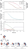
Fig. 6. IsdB:Hb interaction followed by TR-XSS and spectroscopic techniques.
Global fitting (black dashed lines) as a multiexponential function of experimental (panel a and b) and Simbiology-generated (panel c) data. a first (pink) and second (brown) SVD components from absorption spectroscopy. b Fluorescence (light blue) signal. c Simbiology-generated species concentrations: IsdB (green), Hb (red), 1IsdB:βHbtet complex (blue), 2IsdB:βHbtet complex (orange), 1IsdB:Hbdim complex (gray), and 2IsdB:Hbdim complex (purple). d Kinetic model used for the global fitting analysis.

Similar articles

Cited by

References

    1. Lowy, F. D. How Staphylococcus aureus adapts to its host. N. Engl. J. Med.364, 1987–1990 (2011). - PubMed
    1. Mazmanian, S. K. et al. Passage of heme-iron across the envelope of Staphylococcus aureus. Science299, 906–909 (2003). - PubMed
    1. Torres, V. J. et al. Staphylococcus aureus IsdB is a hemoglobin receptor required for heme iron utilization. J. Bacteriol.188, 8421–8429 (2006). - PMC - PubMed
    1. Pishchany, G. et al. IsdB-dependent hemoglobin binding is required for acquisition of heme by Staphylococcus aureus. J. Infect. Dis.209, 1764–1772 (2014). - PMC - PubMed
    1. Zhu, H. et al. Non-heme-binding domains and segments of the Staphylococcus aureus IsdB protein critically contribute to the kinetics and equilibrium of heme acquisition from methemoglobin. PLoS One9, e100744 (2014). - PMC - PubMed

LinkOut - more resources