. 2025 Oct;24(10):1653-1663.
doi: 10.1038/s41563-024-02114-5. Epub 2025 Feb 17.

Blood-brain-barrier-crossing lipid nanoparticles for mRNA delivery to the central nervous system

Affiliations

Blood-brain-barrier-crossing lipid nanoparticles for mRNA delivery to the central nervous system

Chang Wang et al. Nat Mater. 2025 Oct.

Abstract

The systemic delivery of mRNA molecules to the central nervous system is challenging as they need to cross the blood-brain barrier (BBB) to reach into the brain. Here we design and synthesize 72 BBB-crossing lipids fabricated by conjugating BBB-crossing modules and amino lipids, and use them to assemble BBB-crossing lipid nanoparticles for mRNA delivery. Screening and structure optimization studies resulted in a lead formulation that has substantially higher mRNA delivery efficiency into the brain than those exhibited by FDA-approved lipid nanoparticles. Studies in distinct mouse models show that these BBB-crossing lipid nanoparticles can transfect neurons and astrocytes of the whole brain after intravenous injections, being well tolerated across several dosage regimens. Moreover, these nanoparticles can deliver mRNA to human brain ex vivo samples. Overall, these BBB-crossing lipid nanoparticles deliver mRNA to neurons and astrocytes in broad brain regions, thereby being a promising platform to treat a range of central nervous system diseases.


Conflict of interest statement

Competing interests: Y.D. is a scientific advisor in Arbor Biotechnologies, Sirnagen Therapeutics and Moonwalk Biosciences, and also a co-founder and holds equity in Immunanoengineering Therapeutics. J.P. is a current employee of Biogen with salary and stock options. P.C.P. is a current employee of City Therapeutics with salary and stock options. The other authors declare no competing interests.

Figures
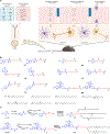
Fig. 1 |
Fig. 1 |. Design of BLNPs.
a, Illustration of the formulation of BLNPs and potential BBB-crossing mechanisms. b, Chemical structures of BLs. c, Synthesis routes of representative BLs (DS11 and MK6).
Fig. 2 |
Fig. 2 |. Characterizations of BLNPs for mRNA delivery.
af, Luminescence intensity of BLNP-FLuc mRNA in N2a cells. The intensity was normalized to the MC3 LNP group. g, Luminescence intensity of brains from the lead BLNP-FLuc-mRNA-treated mice (intravenous). The intensity was normalized to the MC3 LNP group. h, Representative images of brains from the mice intravenously treated with MC3 LNPs and MK6 BLNPs. i, Luminescence quantification of the organs from mice treated with PBS, free mRNA, MC3 LNP or MK6 BLNP. Data in ai are from n = 3 biologically independent samples. Data are presented as mean ± s.d. Statistical significance was determined by one-way ANOVA followed by Dunnett’s multiple comparisons test. n.s., not significant, P > 0.05, *P < 0.05, **P < 0.01, ****P < 0.0001.
Fig. 3 |
Fig. 3 |. Optimization and characterization of MK6 BLNP series for systemic mRNA delivery to mouse brain.
a,b, Normalized luminescence intensity (a) and representative IVIS images (b) of brains of mice intravenously injected with MK6 BLNPs and MK6E BLNPs encapsulating FLuc-mRNA. The intensity was normalized to the MK6 BLNP group. c, Structure optimization of MK6 BLs. d,e, Normalized luminescence intensity (d) and representative images (e) of brains after intravenous injections with MC3 LNPs, MK6E BLNPs or MK16 BLNPs. The intensity was normalized to the MC3 LNP group. f, Immunofluorescence flow cytometry analysis of GFP expressions in different brain cell types from brains of the mice intravenously injected with MK16 BLNP-GFP mRNA or MC3 LNP-GFP mRNA. g, Representative histograms of GFP expression in different brain cell types after various doses of MK16-mediated GFP mRNA delivery (1 mg kg−1) from f. h, Illustration of a bEnd.3-N2a transwell cell assay for BBB penetration assessment. The lower compartment was seeded with N2a cells. i, Luminescence intensity of N2a cells in the lower compartment after MK16 BLNP-FLuc mRNA treatment to the bEnd.3 cells in the upper compartment. The bEnd.3 cells were pretreated with either MβCD or NGST. j,k, Fluorescence intensity (j) and representative IVIS images (k) of the brains of the mice after intravenous injections with MK16 BLNPs encapsulating Alexa Fluor 647-labelled RNAs. The mice were pretreated with either MβCD or NGST. l, Illustration of the proposed BBB-crossing mechanisms of MK16 BLNPs. All data are from n = 3 biologically independent samples and are presented as mean ± s.d. Statistical significance and P values were determined by one-way ANOVA followed by Dunnett’s multiple comparisons test. n.s., not significant, P > 0.05, **P < 0.01, ***P < 0.001, ****P < 0.0001.
Fig. 4 |
Fig. 4 |. MK16 BLNPs for Cre mRNA delivery in the Ai14 mouse model.
a, Diagram depicting that delivery of Cre recombinase mRNA turns on tdTomato expression in Ai14 mice. b, Representative brain images of Ai14 mice intravenously injected with PBS, MK16 BLNP-Cre mRNA or MC3 LNP-Cre mRNA. Scale bar, 1 mm. ce, tdTomato expression in neurons (Map2+) and astrocytes (GFAP+) in the hippocampus (c), thalamus (d) and cortex (e). Scale bar, 50 μm. fh, Quantification of tdTomato-positive neurons and astrocytes in the hippocampus (f), thalamus (g) and cortex (h). i, Immunofluorescence flow cytometry analysis of tdTomato expression in different brain cell types after single or triple intravenous injections of MK16 BLNP-Cre mRNA and MC3 LNP-Cre mRNA. All data are from n = 3 biologically independent samples and are presented as mean ± s.d. Statistical significance and P values were determined by one-way ANOVA followed by Dunnett’s multiple comparisons test. n.s., not significant, P > 0.05, **P < 0.01, ***P < 0.001, ****P < 0.0001.
Fig. 5 |
Fig. 5 |. MK16 BLNPs for ΔFosb mRNA delivery in a CPP model.
a, ΔFOSB expression in the NAc region of the mouse brain after the intravenous administration of MK16 BLNP-ΔFosb mRNA. Scale bar, 100 μm. n = 3 biologically independent samples. b, Schematic depicting the CPP procedure. S, saline; C, cocaine. c, Preference score calculated as time spent on the drug-paired side – time spent in the saline-paired side of the conditioned mice treated with PBS or MK16 BLNP-ΔFosb mRNA. n = 19 mice for the PBS group and n = 20 mice for the MK16 BLNP group. d, Schematic depicting ex vivo mRNA delivery in human brain tissue. IFCM, immunofluorescence flow cytometry. e, ΔFOSB expression levels across neurons, astrocytes and microglia from adult human cerebral cortex dissections after ex vivo treatment with PBS or MK16 BLNP-ΔFosb mRNA. n = 4 tissue slices for each group. Data in c and e are presented as the mean ± s.d. Statistical significance and P values in c were determined by two-way ANOVA with Šidák post hoc test. Statistical significance and P values in e were determined by a two-tailed Student’s t-test. n.s., not significant, P > 0.05, **P < 0.01, ***P < 0.001.
Fig. 6 |
Fig. 6 |. MK16 BLNP-Pten mRNA treatment in an orthotopic GBM mouse model.
a, Schematic of the treatment regimen in the U-118MG GBM model. The treatments were intravenously injected into the tumour-bearing mice via the tail vein at an mRNA dose of 1.0 mg kg−1. A total of three treatments were given at three-day intervals. b, Luminescence intensity of orthotopic GBM tumour tissues in the mice treated with PBS, MK16 BLNP-control mRNA and MK16 BLNP-Pten mRNA via the tail vein. c, Representative IVIS images of the tumour-bearing mice. d, Mouse survival over time. n = 9 mice for the PBS group and control mRNA group. n = 10 mice for the Pten mRNA group. Data in b are presented as the mean ± s.d. Statistical significance and P values in b were determined by two-way ANOVA with Fisher’s least significant difference. Statistical significance and P values in d were analysed by the log-rank (Mantel–Cox) test. *P < 0.05, **P < 0.01, ***P < 0.001, ****P < 0.0001.

References

    1. Weissman D & Karikó K mRNA: fulfilling the promise of gene therapy. Mol. Ther 23, 1416–1417 (2015). - PMC - PubMed
    1. Pardi N, Hogan MJ, Porter FW & Weissman D mRNA vaccines–a new era in vaccinology. Nat. Rev. Drug Discov 17, 261–279 (2018). - PMC - PubMed
    1. Hajj KA & Whitehead KA Tools for translation: non-viral materials for therapeutic mRNA delivery. Nat. Rev. Mater 2, 17056 (2017).
    1. Hou X, Zaks T, Langer R & Dong Y Lipid nanoparticles for mRNA delivery. Nat. Rev. Mater 6, 1078–1094 (2021). - PMC - PubMed
    1. Chaudhary N, Weissman D & Whitehead KA mRNA vaccines for infectious diseases: principles, delivery and clinical translation. Nat. Rev. Drug Discov 20, 817–838 (2021). - PMC - PubMed

LinkOut - more resources