

Baseline choroidal microvasculature dropout as a predictor of rapid global structural loss in open-angle glaucoma
- PMID: 39962316
- PMCID: PMC11832928
- DOI: 10.1038/s41598-025-90198-7
Baseline choroidal microvasculature dropout as a predictor of rapid global structural loss in open-angle glaucoma
Abstract
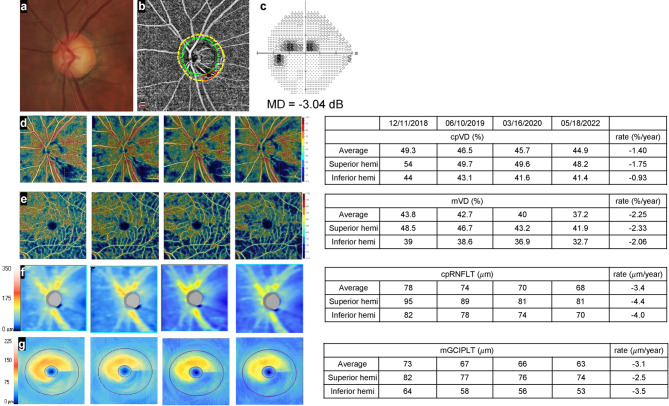
This study included 102 open-angle glaucoma (OAG) eyes with or without a localized choroidal microvasculature dropout (CMvD) at the inferior hemiretina, matched for age (≤ 10 years), axial length (≤ 1 mm), and visual field severity (≤ 1dB), and with a minimum 2-year follow-up. Serial thickness [circumpapillary retinal nerve fiber layer (cpRNFLT) and macular ganglion cell-inner plexiform layer thickness (mGCIPLT)], and vessel density (VD) [circumpapillary (cpVD) and macular VD (mVD)] parameters were obtained. The rate of change in each parameter at both the superior (CMvD-unaffected) and inferior (CMvD-affected) hemiretina were compared between matched eyes with (CMvD+) and without CMvD (CMvD-). Clinical factors associated with the rate of change in each parameter both globally and at the CMvD-unaffected hemiretina were also evaluated. CMvD + eyes showed significantly faster rates of VD and thickness loss at both the CMvD-affected and -unaffected hemiretina. In addition, CMvD was significantly associated with rapid loss of both VD and thickness parameters globally and at the CMvD-unaffected superior hemiretina. In conclusion, OAG eyes with CMvD show significantly faster rates of VD and thickness loss at both the CMvD-affected and unaffected hemiretina. A localized CMvD is an independent predictor of globally rapid structural loss in OAG eyes.
Keywords: Choroidal microvasculature dropout; Glaucoma; Optical coherence tomography angiography; Progression.
© 2025. The Author(s).
Conflict of interest statement
Declarations. Competing interests: The authors declare no competing interests.
Figures


References
-
- Van Buskirk, E. M. & Cioffi, G. A. Glaucomatous optic neuropathy. Am. J. Ophthalmol.113, 447–452. 10.1016/s0002-9394(14)76171-9 (1992). - PubMed
-
- Flammer, J. et al. The impact of ocular blood flow in glaucoma. Prog Retin Eye Res.21, 359–393. 10.1016/s1350-9462(02)00008-3 (2002). - PubMed
-
- Anderson, D. R. & Braverman, S. Reevaluation of the optic disk vasculature. Am. J. Ophthalmol.82, 165–174. 10.1016/0002-9394(76)90414-1 (1976). - PubMed
-
- Onda, E., Cioffi, G. A., Bacon, D. R. & Van Buskirk, E. M. Microvasculature of the human optic nerve. Am. J. Ophthalmol.120, 92–102. 10.1016/s0002-9394(14)73763-8 (1995). - PubMed
-
- Yin, Z. Q., Vaegan, Millar, T. J., Beaumont, P. & Sarks, S. Widespread choroidal insufficiency in primary open-angle glaucoma. J. Glaucoma6, 23–32 (1997). - PubMed
MeSH terms
LinkOut - more resources
Full Text Sources
