. 2025 Feb 17;23(1):114.
doi: 10.1186/s12951-025-03176-6.

RGD hydrogel-loaded ADSC extracellular vesicles mitigate uranium-induced renal injury via TLR4/NF-κB pathway inhibition

Affiliations

RGD hydrogel-loaded ADSC extracellular vesicles mitigate uranium-induced renal injury via TLR4/NF-κB pathway inhibition

Xi Chen et al. J Nanobiotechnology. .

Abstract

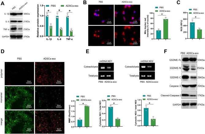
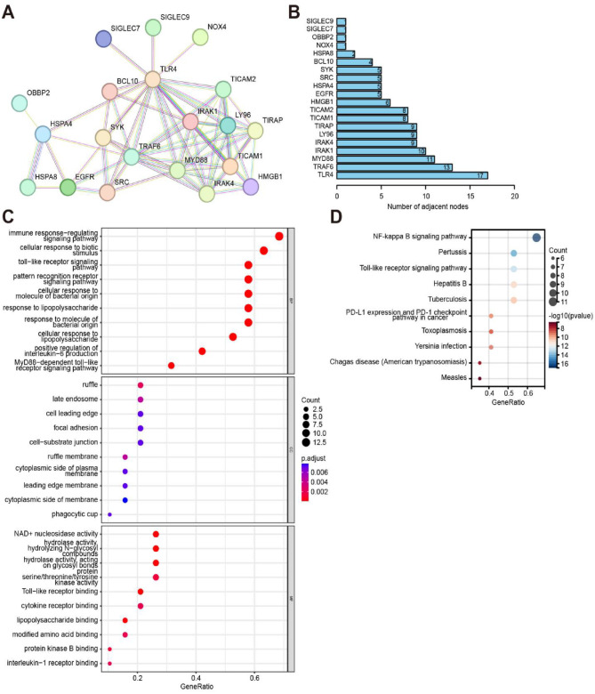
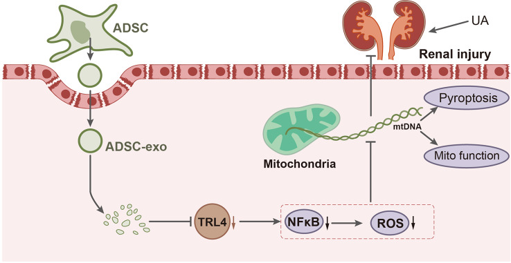
Background: Uranium-induced kidney damage represents a major health concern due to its toxic effects, including mitochondrial dysfunction and inflammation. Mitochondrial DNA (mtDNA)-mediated pyroptosis is a critical pathway in the pathogenesis of renal injury. The toll-like receptor 4 / nuclear factor-kappa B (TLR4/NF-κB) signaling pathway plays a pivotal role in this process. Recent studies have shown that extracellular vesicles derived from adipose-derived stem cells (ADSCs-EVs) possess therapeutic potential due to their anti-inflammatory and regenerative properties. Incorporating ADSCs-EVs into arginine-glycine-aspartate (RGD), hydrogels may enhance their stability and therapeutic efficacy in vivo. This study aims explore the molecular mechanism by which RGD hydrogel-loaded ADSCs-EVs modulate mtDNA-mediated pyroptosis by suppressing the TLR4/NF-κB signaling pathway to alleviate uranium-induced kidney injury.

Results: Repairing mitochondrial dysfunction was found to mitigate mtDNA leakage, thereby inhibiting renal pyroptosis. ADSCs-EVs alleviated uranium-induced renal cell damage by suppressing the TLR4/NF-κB signaling pathway. In vivo animal experiments confirmed that RGD hydrogel-loaded ADSCs-EVs enhanced their stability in the body and improved their therapeutic efficacy against kidney injury.

Conclusion: Our findings reveal that RGD hydrogel-loaded ADSCs-EVs effectively inhibit the TLR4/NF-κB signaling pathway, preventing mtDNA-mediated pyroptosis and alleviating uranium-induced kidney damage. This elucidation provides a novel strategy for utilizing RGD hydrogel-loaded ADSCs-EVs in treating kidney injury.

Keywords: ADSCs-EVs extracellular vesicles; Kidney injury; Mitochondrial DNA; Pyroptosis; RGD hydrogel; TLR4/NF-κB signaling pathway.


Conflict of interest statement

Declarations. Ethical approval: All animal experiments were approved by our hospital’s animal ethics committee (No. 22-07-127) and conformed to ethical norms. Competing interests: The authors declare no competing interests.

Figures

Fig. 1
Fig. 1
Mitochondrial damage and mtDNA release in uranium-induced renal cell injury model. (A) LDH levels in the supernatant of HK-2 cells detected by ELISA; (B) Mitochondrial morphology in Control and UA groups stained with Mitotracker (red, mitochondria; blue, nucleus; scale bar = 15 μm); (C) Total ROS content in Control and UA group cells detected by DCFH-DA assay; (D) MMP in Control and UA groups detected by JC-1 staining, scale bar = 50 μm; (E) Cytoplasmic mtDNA copy number in renal cells after UA treatment detected by qPCR. All cellular experiments were repeated three times, * indicates P < 0.05
Fig. 2
Fig. 2
Mitochondrial damage in uranium-induced kidney injury model. (A) Histological examination of rat kidney tissue morphology by H&E staining (scale bar = 50 μm); (B) Assessment of levels of urea, creatinine, malondialdehyde, glutathione, and superoxide dismutase in rat kidney tissue; (C) Measurement of mRNA levels of relevant cellular factors TWNK, TFAM, MRC I, MRC IV, PGC-1α, NRF1, and NRF2 in rat kidney tissue by RT-qPCR; (D) Protein levels of TWNK, TFAM, PGC-1α, NRF1, and NRF2 were detected via Western blot across different experimental groups; (E) TEM images displaying mitochondria in kidney tissues along with quantitative data on mitochondrial length and cristae density in each group. Scale bars = 1 μm / 500 nm. Each group consisted of 10 rats, * denotes P < 0.05
Fig. 3
Fig. 3
The impact of restoring mitochondrial dysfunction on mtDNA leakage and renal cell pyroptosis. (A) Expression of IL-1β, IL-6, and TNF-α in each group of cells detected by Western blot; (B) Mitotracker staining to examine mitochondrial morphology in each group of cells (red for mitochondria, blue for cell nuclei; scale bar = 15 μm); (C) DCFH-DA reagent used to measure total ROS levels in each group of cells; (D) JC-1 staining to assess MMP in each group of cells, scale bar = 50 μm; (E) Quantitative PCR to determine cytoplasmic mtDNA copy numbers in each group of cells; (F) Expression of activated GSDME and Caspase-3 in each group of cells detected by Western blot. All cell experiments were performed in triplicate, with * indicating P < 0.05
Fig. 4
Fig. 4
ADSCs-EVs alleviate uranium-induced renal cell damage. (A) Western blot analysis was performed to assess the expression of IL-1β, IL-6, and TNF-α in each group of cells; (B) Mitotracker staining was used to examine the mitochondrial morphology of cells in each group (red, mitochondria; blue, nucleus; scale bar = 15 μm); (C) DCFH-DA reagent was employed to measure the total ROS levels in each group of cells; (D) JC-1 staining was conducted to evaluate the MMP of cells in each group, with a scale bar of 50 μm; (E) RT-qPCR was utilized to determine the cytoplasmic mtDNA copy number in each group of cells; (F) Western blot analysis was carried out to determine the expression of activated GSDME and Caspase-3 in each group of cells. All cell experiments were performed in triplicate, and * indicates P < 0.05
Fig. 5
Fig. 5
Possible mechanism of ADSCs-EVs inhibition of mtDNA-mediated cell apoptosis. (A) PPI network constructed by obtaining proteins interacting with TLR4 from the STRING database; (B) Sorting of the number of adjacent nodes in the PPI network representing protein interactions; (C) Functional enrichment analysis of proteins in the PPI network using GO; (D) Enrichment analysis of KEGG pathways for proteins in the PPI network
Fig. 6
Fig. 6
Effect of ADSCs-EVs on uranium-induced renal cell injury via modulation of TLR4/NF-κB signaling pathway figure X. Overview of experimental procedures. (A) Western blot to assess TLR4 expression levels in various cell groups; (B) Western blot to detect nuclear expression of p-p65 in different cell groups; (C) Immunofluorescence analysis to examine nuclear translocation of p65 in various cell groups; (D) Western blot after PMA treatment to measure nuclear expression of p-p65 in different cell groups; (E) Immunofluorescence analysis post-PMA treatment to evaluate nuclear translocation of p65 in different cell groups; (F) Western blot for IL-1β, IL-6, and TNF-α expression in various cell groups; (G) Mitotracker staining to assess mitochondrial morphology in different cell groups (red, mitochondria; blue, nucleus; scale bar = 15 μm); (H) DCFH-DA assay to measure total ROS content in various cell groups; (I) JC-1 staining to evaluate MMP in different cell groups, scale bar = 50 μm; (J) RT-qPCR to determine cytoplasmic mtDNA copy number in various cell groups; (K) Western blot for activated GSDME and Caspase-3 expression in different cell groups. All cell experiments were performed in triplicate, with * indicating P < 0.05
Fig. 7
Fig. 7
Effect of RGD hydrogel-loaded ADSCs-EVs on the in vivo stability of ADSCs-EVs. (A) TEM image shows intertwined nanofibers; (B) Schematic illustration of RGD hydrogel-loaded ADSCs-EVs; (C) Dynamic frequency scans of ADSCs-EVs/RGD hydrogel encapsulating ADSCs-EVs at different mass ratios (0.5:1 (green), 1:1 (blue), 1.5:1 (pink), 2:1 (yellow)), where G’ represents storage modulus and G’’ represents loss modulus; (D) Dynamic frequency scans of RGD hydrogel encapsulating ADSCs-EVs at a 1:1 mass ratio before (black) and after (blue) encapsulation, showing G’ and G’’ values; (E) In vivo retention time of Cy5-labeled RGD hydrogel; (F) Bioluminescence imaging of Gluc-labeled ADSCs-EVs indicating a linear dependency between ADSCs-EVs concentration and Gluc signal; (G) Bioluminescence imaging tracking the in vivo stability of ADSCs-EVs. * indicates P < 0.05
Fig. 8
Fig. 8
Therapeutic Effects of RGD Hydrogel-Loaded ADSCs-EVs on Kidney Injury. (A-B) Immunofluorescence staining showing the colocalization of p65 with the cell nucleus in rat renal tissues of each group; (C) H&E staining examining the morphological structure of rat kidney tissues in each group (scale bar = 50 μm); (D) Assessment of urea, creatinine, malondialdehyde, glutathione, and superoxide dismutase levels in rat kidney tissues of each group; (E) RT-qPCR analysis of mRNA levels of relevant cytokines TWNK, TFAM, MRC I, MRC IV, PGC-1α, NRF1, and NRF2 in rat kidney tissues of each group; (F) TEM images displaying mitochondria in kidney tissues in each group, along with quantitative data on mitochondrial length and cristae density, Scale bars = 1 μm / 500 nm. Each group consisted of 10 rats, with * indicating P < 0.05
Fig. 9
Fig. 9
ADSCs-EVs alleviate uranium-induced kidney injury by inhibiting the TLR4/NF-κB signaling pathway

References

    1. Ortega-Romero M, Jiménez-Córdova MI, Barrera-Hernández Á, et al. Relationship between urinary biomarkers of early kidney damage and exposure to inorganic toxins in a pediatric population of Apizaco, Tlaxcala, Mexico. J Nephrol. 2023;36(5):1383–93. 10.1007/s40620-023-01598-9. - PubMed
    1. Al-Full ZZ, Khattab MR. Uranium isotopic ratio of black shale and its role in detection of oxic-anoxic conditions of uranium depositions. J Environ Sci Health Tox Hazard Subst Environ Eng. 2022;57(5):376–85. 10.1080/10934529.2022.2068886. - PubMed
    1. Xue XM, Wu XY, Zhan JM. Zhonghua Lao Dong Wei Sheng Zhi Ye Bing Za Zhi. 2023;41(11):864–9. 10.3760/cma.j.cn121094-20221209-00589 - PubMed
    1. Zhong D, Wang R, Zhang H, Wang M, Zhang X, Chen H. Induction of lysosomal exocytosis and biogenesis via TRPML1 activation for the treatment of uranium-induced nephrotoxicity. Nat Commun. 2023;14(1):3997. Published 2023 Jul 6. 10.1038/s41467-023-39716-7 - PMC - PubMed
    1. Su SH, Su SJ, Huang LY, Chiang YC. Leukemic cells resist lysosomal inhibition through the mitochondria-dependent reduction of intracellular pH and oxidants. Free Radic Biol Med. 2023;198:1–11. 10.1016/j.freeradbiomed.2023.01.025. - PubMed

MeSH terms

LinkOut - more resources