. 2025 Feb 17;23(1):113.
doi: 10.1186/s12951-025-03196-2.

Intraperitoneal administration of mRNA encoding interleukin-12 for immunotherapy in peritoneal carcinomatosis

Affiliations

Intraperitoneal administration of mRNA encoding interleukin-12 for immunotherapy in peritoneal carcinomatosis

Leire Arrizabalaga et al. J Nanobiotechnology. .

Abstract

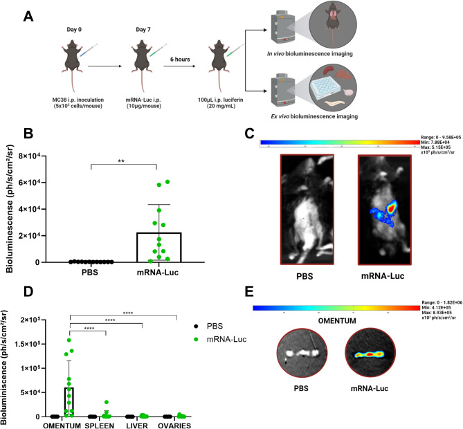
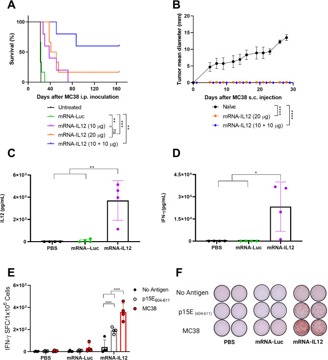
Peritoneal carcinomatosis is an advanced stage of cancer with very limited treatment options. Locoregional immunotherapy is being evaluated as a way to improve efficacy and limit toxicity. This study assessed the efficacy of a cationic polymer/lipid-based transfection compound in delivering mRNA molecules intraperitoneally. Our investigation of the transfer of luciferase mRNA in murine models of peritoneal carcinomatosis revealed preferential luciferase expression in the omentum upon the intraperitoneal administration of complexed mRNAs. Macrophages were identified as key cells that capture and express the mRNA complexes, and accordingly, depletion of resident macrophages led to reduced reporter luciferase expression. To explore the therapeutic potential of this approach, mRNA complexes encoding single-chain interleukin-12 (IL12), an immunostimulatory molecule (mRNA-IL12), were investigated. mRNA-IL12-treated mice exhibited a significant survival advantage in models of peritoneal carcinomatosis and acquired immune memory, as shown upon subcutaneous rechallenge. Tumor microenvironment analyses revealed increased numbers of CD4+ and CD8+ T cells with a more proliferative phenotype, accompanied by decreased myeloid populations in the omentum. Overall, our study underscores the potential of mRNA complexes for efficient mRNA delivery, eliciting effective antitumor responses and modulating the tumor microenvironment to treat peritoneal carcinomatosis.

Keywords: Cancer immunotherapy; Interleukin-12; Locoregional treatment; Peritoneal carcinomatosis; mRNA.


Conflict of interest statement

Declarations. Ethics approval and consent to participate: All the experiments received approval (R-080–19GN) from the Ethics Committee for Animal Testing at the University of Navarra. Consent for publication: Not applicable. Competing interests: Ignacio Melero reports receiving commercial research grants from AstraZeneca, BMS, Highlight Therapeutics, Alligator, Pfizer Genmab and Roche; has received speakers bureau honoraria from MSD; and is a consultant or advisory board member for BMS, Roche, AstraZeneca, Genmab, Pharmamar, F-Star, Bioncotech, Bayer, Numab, Pieris, Gossamer, Alligator and Merck Seron.The rest of the authors report no conflicts of interest in this work.

Figures

Fig. 1
Fig. 1
Intraperitoneal administration of mRNA complexes allows expression of the encoded protein preferentially in omentum (A) The scheme summarizes the experimental design. A‒E. MC38 tumor-bearing C57BL/6 mice (n = 9‒12/group) received one dose of mRNA-Luc (10 µg of luciferase-encoding mRNA complexes) or PBS (control group) i.p. 7 days after tumor inoculation. Six hours later, bioluminescence intensity was measured via PhotonIMAGER in vivo (B-C) and ex vivo (D-E). (B) In vivo bioluminescence quantification (ph/s/cm2/sr) 6 h after PBS or mRNA-Luc i.p. administration. (C) Representative image of luciferase expression in vivo. (D) Six hours after PBS or mRNA-Luc i.p. administration, the omentum, spleen, liver and ovaries were collected, and ex vivo bioluminescence was quantified (ph/s/cm2/sr). (E) Image illustrating the expression of luciferase in the omentum. Student’s t tests were performed in panel B. Two-way ANOVA followed by Sidak’s multiple comparison test for panel D. **p value < 0.01, ****p value < 0.0001
Fig. 2
Fig. 2
Clodronate-depletable macrophages in the peritoneal cavity are involved in the capture and expression of mRNA complexes. (A) C57BL/6 mice were challenged i.p. with 5 × 105 MC38 colon cancer cells. Six days later, the mice were treated i.p. with PBS (control group) or 200 µl of clodronate liposome solution (5 mg/ml). One day later, mRNA-Luc (10 µg/mouse) was administered i.p., and bioluminescence intensity was measured via PhotonIMAGER in vivo and ex vivo 6 h later (n = 6/group). (B) Six hours after PBS or mRNA-Luc i.p. injection, luciferase expression was quantified (ph/s/cm2/sr) in vivo. Six mice were treated with clodronate liposomes only as a control group. (C) Representative image of luciferase expression in vivo in mice treated with mRNA-Luc alone or with the combination of clodronate liposomes and mRNA-Luc. (D) Ex vivo bioluminescence measurement (ph/s/cm2/sr) of the omentum, spleen, liver and ovaries 6 h after mRNA-Luc administration i.p. (E) Image showing luciferase expression in the omentum. One-way ANOVA for panel B and two-way ANOVA followed by Tukey’s multiple comparison test for panel D. *p value < 0.05, ****p value < 0.0001
Fig. 3
Fig. 3
IL12-encoded mRNA complexes exert antitumor effects on MC38-derived peritoneal carcinomatosis. (A) Survival of 5 × 105 MC38 i.p. tumor-bearing mice (n = 5–6/group) treated twice with PBS or mRNA-IL12 (10 µg/mouse) at days 10 and 13 or once with mRNA-Luc (20 µg/mouse), mRNA-IL12 (10 µg/mouse) or mRNA-IL12 (20 µg/mouse) on day 10 after tumor challenge. (B) MC38 cells (5 × 105) were injected subcutaneously into the mice 165 days after primary tumor challenge. Six naïve mice were used as a control group. The tumor follow-up data are shown. CF. MC38 cells (5 × 105) were administered i.p. to C57BL/6 mice (n = 4/group). The mice were treated i.p. twice with PBS, mRNA-Luc or mRNA-IL12 (10 µg/mouse) at days 10 and 13 after tumor inoculation. Eighteen hours after the second treatment, the mice were sacrificed, and spleen and peritoneal lavage samples were collected for further analysis. C-D. ELISAs were carried out with peritoneal lavage samples to detect IL12 and IFN-γ. E. Splenocytes were stimulated with p15E604 − 611 antigen or with 1 × 105 irradiated MC38 tumor cells (20,000 rads), and the number of IFN-γ–producing cells was measured via ELISpot. F. A representative ELISpot plate image. Log-rank (Mantel‒Cox) tests were used to analyze the survival data in panel A. The data from panel B are expressed as the means ± SDs, and repeated-measures ANOVA was used for the statistical analysis. Statistical significance was determined with one-way ANOVA followed by Tukey’s multiple comparisons tests in panels C and D. In panel E, two-way ANOVA followed by Tukey’s multiple comparisons test was performed. *p < 0.05, **p value < 0.01, ***p < 0.001, ****p < 0.0001
Fig. 4
Fig. 4
Intraperitoneal administration of IL12-encoding mRNA complexes results in a potent antitumor response in peritoneal Panc02.OVA peritoneal carcinomatosis model. A-G. Panc02.OVA cells (5 × 105) were injected i.p. into C57BL/6 mice. Ten days later, 10 µg of IL12 mRNAs were delivered with cationic/polymer complexes in 100 µl of free DMEM i.p. BC. Sera were collected from the mice at the indicated time points, and ELISAs were performed to detect IL12 and IFN-γ (n = 4/group). C. Schematic representation of the experimental design and survival follow-up of mice treated with PBS, mRNA-Luc or mRNA-IL12 i.p. 10 days after tumor challenge (n = 10/group). D. Six days after treatment, the mice were sacrificed (n = 6/group), and an ELISpot assay was used to measure the number of IFN-γ–producing splenocytes stimulated with 4 × 104 irradiated Panc02.OVA cells (20,000 rads). E. Image of two representative wells per condition from the ELISpot plate. The data from panels A-B are expressed as the means ± SDs, and repeated-measures ANOVA was used for the statistical analysis. The survival data in panel C were analyzed via log-rank (Mantel‒Cox) tests. The statistical significance in panel D was determined via two-way ANOVA followed by Tukey’s multiple comparisons test. *p value < 0.05, ****p value < 0.0001.
Fig. 5
Fig. 5
IL12-encoding mRNA complexes favorably modulate the microenvironment in Panc02.OVA peritoneal carcinomatosis model. 5 × 105 Panc02.OVA cells were injected i.p. into C57BL/6 mice (n = 6/group). Ten days later, 10 µg of IL12 mRNAs were delivered with cationic/polymer complexes in 100 µl of free DMEM i.p. Six days after treatment, the mice were sacrificed, and the spleens, peritoneal lavage samples and omentum were collected for further analysis via flow cytometry. Multicolor flow cytometry analysis was used to characterize the immune cells in the samples phenotypically. (A) A dimension-reduced projection of various color-coded cell types from the peritoneal lavage fluid is represented in the UMAP graphics. (B) Abundance boxplot displaying group differences. (C) Lymphoid and myeloid populations from the peritoneal lavage samples were analyzed by flow cytometry. The expression of Ki67 was assessed in CD45+CD19-TCRβ+CD4+ and CD45+CD19−TCRβ+CD8+ cells. (D) Abundance boxplot showing differences between groups. (E) UMAPs illustrating a dimension-reduced projection of different color-coded cell types from the omentum.. (F) Flow cytometry was used to evaluate the lymphoid and myeloid populations from the omentum. Statistical significance was determined with one-way ANOVA followed by Tukey’s multiple comparisons tests. *p < 0.05, **p < 0.001, ***p < 0.001, ****p < 0.0001, ns: nonsignificant

References

    1. Sadeghi B, Arvieux C, Glehen O, et al. Peritoneal carcinomatosis from non-gynecologic malignancies: results of the EVOCAPE 1 multicentric prospective study. Cancer Jan. 2000;15(2):358–63. 10.1002/(sici)1097-0142(20000115)88:2%3C;358::aid-cncr16%3E;3.0.co;2-o - PubMed
    1. Coccolini F, Gheza F, Lotti M, et al. Peritoneal carcinomatosis. World J Gastroenterol Nov. 2013;7(41):6979–94. 10.3748/wjg.v19.i41.6979 - PMC - PubMed
    1. Aranda F, Eguren-Santamaria I, Bella A, Berraondo P, Melero I. Revisiting Intracavitary Immunotherapy of Cancer. Clin Cancer Res May. 2022;13(10):1993–5. 10.1158/1078-0432.CCR-22-0201 - PubMed
    1. Verwaal VJ, van Ruth S, de Bree E, et al. Randomized trial of cytoreduction and hyperthermic intraperitoneal chemotherapy versus systemic chemotherapy and palliative surgery in patients with peritoneal carcinomatosis of colorectal cancer. J Clin Oncol Oct. 2003;15(20):3737–43. 10.1200/JCO.2003.04.187 - PubMed
    1. Cortes-Guiral D, Hubner M, Alyami M, et al. Primary and metastatic peritoneal surface malignancies. Nat Rev Dis Primers Dec. 2021;16(1):91. 10.1038/s41572-021-00326-6 - PubMed