Review
. 2025 Feb 17;24(1):52.
doi: 10.1186/s12944-025-02463-3.

Crosstalk between renin and arachidonic acid (and its metabolites)

Affiliations
Review

Crosstalk between renin and arachidonic acid (and its metabolites)

Undurti N Das et al. Lipids Health Dis. .

Abstract

Renin plays a significant role in the regulation of blood pressure and fluid volume by modulating the renin‒angiotensin‒aldosterone (RAAS) system. Renin suppression reduces serum aldosterone levels and lowers blood pressure in addition to preserving renal function. However, exactly how renin synthesis and action are regulated and how renin suppression preserves renal function are not clear. We propose that arachidonic acid (AA) and its metabolites control renin synthesis, secretion, and action by virtue of its (AA) anti-inflammatory, cytoprotective actions and ability to regulate the secretion of renin. These findings suggest that direct renin suppression results in changes in AA metabolism. This proposal implies that AA and its metabolites may be developed as potential drugs to prevent and manage hypertension and preserve renal function.

Keywords: Arachidonic acid; Inflammation; Neurotransmitters; Nitric oxide; Renin; Renin-angiotensin-aldosterone system.


Conflict of interest statement

Declarations. Ethics approval and consent to participate: Not applicable. Competing interests: The authors declare no competing interests.

Figures

Fig. 1
Fig. 1
The structure of the glomerular apparatus (JGA)
Fig. 2
Fig. 2
Scheme showing the close relationships among the kidney, liver, lungs, adrenal cortex and vascular tissue and their relevance to the renin‒angiotensin‒aldosterone system (RAAS)
Fig. 3
Fig. 3
Scheme showing the potential relationships among prorenin, renin, angiotensin II and EFAs and their metabolites and their relevance to some diseases. Polyunsaturated fatty acids (PUFAs: DGLA, AA, EPA and DHA) and their metabolites, such as PGs, LTs, TXs, LXA4, resolvins, protectins and maresins, can alter (suppress) the activities of renin and angiotensin-II and thus influence renal function, blood pressure, and blood volume. In contrast, PGs, LTs and TXs may increase the activities of renin, ACE, and angiotensin-II. Under physiological conditions, there is a balance between PGs, LTs and TXs vs. LXA4, RSVs, PRTs, and MaRs. PUFAs can alter cell membrane fluidity and thus alter the expression and binding of various growth factors and hormones to their receptors. LXA4 = Lipoxin A4; RSVs = Resolvins; PRTs = Protectins; MaRs = Maresins
Fig. 4
Fig. 4
Scheme showing the Renin and prorenin conformations. Under physiological conditions (pH 7.4, 37 °C), only ≈ 1% or less of prorenin occurs in the so-called ‘open’ conformation, i.e., a conformation where the prosegment has moved out of the enzymatic cleft, thus allowing it to react with angiotensinogen to yield angiotensin I. However, the majority of prorenin is closed and inactive. This ‘proteolytic’ activation occurs exclusively in the kidney, and the enzyme responsible for this activation is not yet known
Fig. 5
Fig. 5
Scheme showing the changes in various hormones starting from the luteal phase of the menstrual cycle, during pregnancy and during the postpartum period. These chnages include systemic vasodilation; a decrease in peripheral vascular resistance; a reduction in maternal systolic, diastolic and mean arterial blood pressure; and a return to nonpregnant levels during the last month of pregnancy
Fig. 6
Fig. 6
Scheme showing the interaction of multiple factors secreted by the corpus luteum and their modulatory effects on the RAAS in human pregnancy. Progesterone acts as a mineralocorticoid receptor antagonist and thus blocks the effect of aldosterone. Ang. = angiotensin; AT1R = angiotensin-II type 1 receptor; AT2R = angiotensin-II type 2 receptor; GFR = glomerular filtration rate; MAP = mean arterial pressure; Na+ = sodium; NO = nitric oxide; RPF = renal plasma flow
Fig. 7
Fig. 7
Scheme showing local prorenin synthesis in the ovaries, uteroplacental unit and fetal membranes, known stimulators and inhibitors of prorenin synthesis, and possible functions (local and systemic) of prorenin. ATP = adenosine triphosphate; Ca 2 + = extracellular calcium; cAMP = cyclic adenosine monophosphate; hCG = human chorionic gonadotropin; PDE = phosphodiesterase; PDEi = phosphodiesterase inhibitor. PKC = protein kinase C; TNF = tumor necrosis factor; VEGF = vascular endothelial growth factor
Fig. 8
Fig. 8
Scheme showing how AA functions as a mechanotransducer and regulates renin synthesis and release. ECM = Extracellular matrix AA = Arachidonic acid; PGE2 = prostaglandin E2; LXA4 = lipoxin A4; NF-kB = Nuclear factor-kappa B; IL-6 = Interleukin-6; TNF = Tumor necrosis factor; HPETE = Hydroperoxyeicosatetraenoic acid; HETE = Hydroxyeicosatetraenoic acid; CREB = cAMP-binding protein. AA itself or its metabolites act on F-actin, lamin A/C and chromatin to regulate renin gene expression. Metabolites of AA, PGE2, LXA4, HPETE and HETE can either increase or decrease renin formation in response to changes in perfusion pressure. PGE2 can either increase or decrease renin synthesis, depending on its concentration, and bind to its various receptors. When PGE2 reaches its optimum level, it triggers the synthesis of LXA4 from AA and LXA4, which in turn inhibits PGE2 formation. NF-kB, angiotensin-II, aldosterone, IL-6 and TNF-α stimulate PLA2 to induce the release of AA from the cell membrane and enhance the formation of PGE2 and other metabolites of AA. LXA4 suppresses the expression of NF-kB and inhibits the synthesis of IL-6 and TNF-α. HPETE and HETE are proinflammatory molecules formed from AA that inhibit renin synthesis. AA and LXA4 increase the synthesis of nitric oxide (NO), a potent vasodilator. This positive and negative feedback regulation among AA, PGE2, LXA4, NF-kB, IL-6, TNF-α, angiotensin-II and cAMP can fine-tune renin synthesis and exocytosis to regulate perfusion pressure and blood pressure. These findings suggest that direct renin suppression enhances the release of AA, which is converted to LXA4 and PGI2, and enhances the formation of NO, which lowers blood pressure and protects renal tissue, thus preserving renal function. Although the emphasis has been on AA and its metabolites, other unsaturated fatty acids, such as GLA (gamma-linolenic acid), DGLA (dihomo-GLA), EPA and DHA, may also have similar beneficial effects. This figure has been modified from reference no. 24
Fig. 9
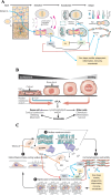
Fig. 9
A The nucleus acts as an elastic mechanotransducer of cellular shape and controls the dynamic behavior of the cell. In response to pressure, the cell shape changes, leading to inner nuclear membrane unfolding and the activation of the cPLA2-AA pathway. AA is the precursor of various eicosanoids that have several physiological and pathological actions. The unfolding of the inner nuclear membrane transduces myosin II to the cell cortex, where it regulates actin cytoskeleton contractility, which results in cell motility as needed. B Nuclear membrane transduction. In response to pressure or stretch stimuli, stretch in the nuclear membrane occurs in conjunction with calcium, which activates cPLA2 release and the release of AA. Eicosanoids formed from AA mediate cell autonomous and paracrine effects. C In response to physical pressure, cell nuclear deformation and unfolding and stretching of the nuclear envelope (2) trigger calcium release, cPLA2 activation and AA release, the precursors of several eicosanoids. These events lead to actomyosin force generation (3) and increased cell migratory capacity (4). A, B and C were created and modified from references 34 and 35
Fig. 10
Fig. 10
A scheme showing the effect of mechanical forces (including blood flow shear stress in endothelial cells, which could be transmitted to renin cells) that modulate the actin–myosin cytoskeletal system and consequently alter the cell shape and eventually lead to chromatin reorganizations and mRNA expression and cell fate
Fig. 11
Fig. 11
Metabolism of essential fatty acids (EFAs)

References

    1. Ueno M, Fujii W, Ono W, Murata H, Fujigaki Y, Shibata S. Renin inhibition and the long-term renal function in patients with 3 hypertensive emergency: a retrospective cohort study. Am J HTN, in press. - PubMed
    1. Heesch CM. Reflexes that Control Cardiovascular function. Am J Physiol Content. 1999;277:S234. - PubMed
    1. Watanabe H, Belyea BC, Paxton RL, Li M, Dzamba BJ, DeSimone DW, Gomez RA, Sequeira-Lopez ML. S. Renin Cell Baroreceptor, a Nuclear Mechanotransducer Central for Homeostasis. Circ Res. 2021;129:262–76. - PMC - PubMed
    1. Schalekamp MADH, Deinum J, Danser AHJ. Short-lived active prorenin: Precursor of So-Called native prorenin. Hypertension. 2024;81(6):1190–3. - PMC - PubMed
    1. Nguyen G, Delarue F, Burcklé C, Bouzhir L, Giller T, Sraer JD. Pivotal role of the renin/prorenin receptor in angiotensin II production and cellular responses to renin. J Clin Invest. 2002;109(11):1417–27. - PMC - PubMed

LinkOut - more resources