Case Reports
. 2025 Feb 10:2025:5548590.
doi: 10.1155/crid/5548590. eCollection 2025.

Oral Squamous Cell Carcinoma in Atrophic-Erosive Lichen Planus: 10-Year Rehabilitative Case Report

Affiliations
Case Reports

Oral Squamous Cell Carcinoma in Atrophic-Erosive Lichen Planus: 10-Year Rehabilitative Case Report

Eduardo Anitua et al. Case Rep Dent. .

Abstract

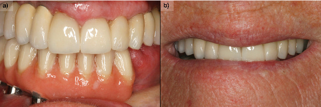
Patients with oral lichen planus can sometimes develop malignancy of the process and develop oral squamous cell carcinoma or another type of cancer. Close monitoring of the lesions and early diagnosis is important to increase patient survival. Once cancer treatment has been performed, the therapeutic options for restoring masticatory and phonatory function, in addition to resolving the aesthetic sequelae, are important. In the present clinical case, we show a patient who was treated and followed over a long period of time for both her carcinoma and her subsequent rehabilitation with implants.


Conflict of interest statement

E.A. is the scientific director of BTI Biotechnology Institute, the company that has developed the Endoret PRGF technology and commercializes BTI implant system. L.P. has no conflict of interest. M.H.A. is a researcher at BTI Biotechnology Institute I MAS D.

Figures

Figure 1
Figure 1
(a) Aspect of the lesion consulted by the patient. We observe the whitish outgrowth areas and how it is introduced under the prosthesis, so we proceed to lift the crowns. (b) X-ray 6 months before the debut of the lesion. (c) Panoramic x-ray on removal of the prosthesis showing bone loss, which may initially suggest peri-implantitis.
Figure 2
Figure 2
Image of the lesion 20 days after waiting for the histological examination results, confirming the presence of oral squamous cell carcinoma. Lateral view shows the lesion extending along the gingival margin.
Figure 3
Figure 3
X-ray of the patient with the osteosynthesis plate at the end of the surgery (6 months after resection surgery).
Figure 4
Figure 4
X-ray with bone consolidation and after removal of the osteosynthesis plate (2.5 years after surgery).
Figure 5
Figure 5
(a) Intraoral images of the patient after surgery with the graft consolidated and healed (6 months after resection surgery). (b) Appearance of the consolidated skin graft on the oral mucosa at higher magnification.
Figure 6
Figure 6
X-ray after placement of the implants and the immediately loaded provisional prosthesis (2.5 years before surgery).
Figure 7
Figure 7
(a) Definitive prosthesis placed in the patient, after 6 months of progressive loading with the initial immediately loaded prosthesis. (b) Image of a smile with the definitive prosthesis.
Figure 8
Figure 8
Panoramic radiograph after placement of the definitive prosthesis with the extension to the third quadrant (3 years after surgical resection).
Figure 9
Figure 9
Implant placed in Position 22 to subsequently extract the remaining teeth and make a complete upper prosthesis with the implants already placed previously. Five years have now passed since the surgery to treat oral squamous cell carcinoma.
Figure 10
Figure 10
(a) Patient at 10 years of follow-up with definitive upper and lower prostheses. (b) Image of the patient's smile at the end of the follow-up.
Figure 11
Figure 11
X-ray at 10 years of follow-up with the stability of both the upper and lower implants, as well as the bone grafted in the mandible as can be seen where it is no longer possible to differentiate the points of union to the original mandible.

References

    1. Al-Hashimi I., Schifter M., Lockhart P. B., et al. Oral lichen planus and oral lichenoid lesions: diagnostic and therapeutic considerations. Oral Surgery, Oral Medicine, Oral Pathology, Oral Radiology, and Endodontology . 2007;103:S25.e1–S25.e12. doi: 10.1016/j.tripleo.2006.11.001. - DOI - PubMed
    1. McCartan B. E., Healy C. M. The reported prevalence of oral lichen planus: a review and critique. Journal of Oral Pathology & Medicine . 2008;37(8):447–453. doi: 10.1111/j.1600-0714.2008.00662.x. - DOI - PubMed
    1. Scully C., El-Kom M. Lichen planus: review and update on pathogenesis. Journal of Oral Pathology . 1985;14(6):431–458. doi: 10.1111/j.1600-0714.1985.tb00516.x. - DOI - PubMed
    1. Roopashree M. R., Gondhalekar R. V., Shashikanth M. C., George J., Thippeswamy S. H., Shukla A. Pathogenesis of oral lichen planus- a review. Journal of Oral Pathology & Medicine . 2010;39(10):729–734. doi: 10.1111/j.1600-0714.2010.00946.x. - DOI - PubMed
    1. George S., John S. A., Anandaraj S., Issac J. S., Harris A., Reshmi J. Childhood oral lichen planus: report of two cases. Journal of Dentistry (Tehran, Iran) . 2015;12(5):374–378. - PMC - PubMed

Publication types

LinkOut - more resources