Comment
. 2024 Dec 17;7(1):fcae453.
doi: 10.1093/braincomms/fcae453. eCollection 2025.

Biallelic NDUFA13 variants lead to a neurodevelopmental phenotype with gradual neurological impairment

Affiliations
Comment

Biallelic NDUFA13 variants lead to a neurodevelopmental phenotype with gradual neurological impairment

Rauan Kaiyrzhanov et al. Brain Commun. .

Abstract

Biallelic variants in NADH (nicotinamide adenine dinucleotide (NAD) + hydrogen (H))-ubiquinone oxidoreductase 1 alpha subcomplex 13 have been linked to mitochondrial complex I deficiency, nuclear type 28, based on three affected individuals from two families. With only two families reported, the clinical and molecular spectrum of NADH-ubiquinone oxidoreductase 1 alpha subcomplex 13-related diseases remains unclear. We report 10 additional affected individuals from nine independent families, identifying four missense variants (including recurrent c.170G > A) and three ultra-rare or novel predicted loss-of-function biallelic variants. Updated clinical-radiological data from previously reported families and a literature review compiling clinical features of all reported patients with isolated complex I deficiency caused by 43 genes encoding complex I subunits and assembly factors are also provided. Our cohort (mean age 7.8 ± 5.4 years; range 2.5-18) predominantly presented a moderate-to-severe neurodevelopmental syndrome with oculomotor abnormalities (84%), spasticity/hypertonia (83%), hypotonia (69%), cerebellar ataxia (66%), movement disorders (58%) and epilepsy (46%). Neuroimaging revealed bilateral symmetric T2 hyperintense substantia nigra lesions (91.6%) and optic nerve atrophy (66.6%). Protein modeling suggests missense variants destabilize a critical junction between the hydrophilic and membrane arms of complex I. Fibroblasts from two patients showed reduced complex I activity and compensatory complex IV activity increase. This study characterizes NADH-ubiquinone oxidoreductase 1 alpha subcomplex 13-related disease in 13 individuals, highlighting genotype-phenotype correlations.

Keywords: Leigh syndrome; NDUFA13; complex I deficiency; mitochondrial disorders; neurodevelopmental disorder.


Conflict of interest statement

D.A.C. and M.N. are employees of GeneDx, LLC.

Figures

Graphical Abstract
Graphical Abstract
Figure 1
Figure 1
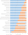
Clinical–radiological features. Clinical features of the affected individuals with biallelic NDUFA13 variants. The bar chart illustrates the most common clinical symptoms and signs associated with NDUFA13-related disease, presented in descending order of frequency. The x-axis represents the total number of patients (N = 13), while the y-axis lists the symptoms and signs. The blue segments indicate the number of patients exhibiting these symptoms/signs, the orange segments show the number of patients without them and the grey segments represent patients for whom the presence or absence of these symptoms/signs is unconfirmed. MRS, magnetic resonance spectroscopy.
Figure 2
Figure 2
Neuroimaging features of NDUFA13-related disease. Most of the individuals present symmetric T2-hyperintense lesions of the substantia nigra (thin white arrows). These lesions are less evident on follow-up scans, as seen in Patient 2b, Patient 7b, and Patient 11b and c and Patient 12b). Periaqueductal and central midbrain signal alterations are visible in individuals Patient 3, Patient 6, Patient 9, Patient 11 and Patient 12 (white arrowheads). In three patients (Patient 2, Patient 3 and Patient 7), there are additional basal ganglia signal alterations (empty white arrows), which increase over time with associated atrophy in individual Patient 2, as depicted in Patient 2b. Faint T2 hyperintensities of the dentate nuclei are noticed in individuals Patient 3, Patient 4, Patient 5, Patient 8 and Patient 12 (thick white arrows). Subtle inferior olivary nuclei T2 hyperintensity is visible in individuals Patient 4 and Patient 11 (dashed black arrows). In one individual (Patient 6), there are T2 signal alterations at the level of the cervical spinal cord (dashed oval). Small foci of restricted diffusion (dashed white arrows) are noted in the right basal ganglia and bilateral substantia nigra (Patient 2a), left periventricular frontal white matter (Patient 6), bilateral substantia nigra (Patient 7a), periaqueductal grey matter (Patient 9) and posterior medullary white matter tracts (Patient 10). Note: mild cerebellar atrophy mainly involving the cerebellar hemispheres in Patients 4 and 11 (curved arrows in Patient 4 and Patient 11c). Pt, patient.
Figure 3
Figure 3
Pedigrees with variant segregation, NDUFA13 variant localizations on gene and protein schematics and variant conservation data. (A) Family trees of the affected individuals with biallelic NDUFA13 variants. Square = male; circle = female; black filled symbol = affected individual; white symbols = unaffected individuals; diagonal line = deceased individual. Double lines indicate consanguinity. The allele with the variant is indicated by the + sign. Wild-type allele is indicated by the −sign. * denotes already published families. (B) Schematic representation of the NDUFA13 gene showing the position of the variants (magenta denotes newly identified variants, blue denotes previously reported variants). (C) Diagram of the NDUFA13 protein with the position of the NDUFA13 variants. Numbers indicate amino acids. (D) Interspecies alignment performed with Clustal Omega shows the level of conservation down to invertebrates on the missense NDUFA13 variants. TM, transmembrane.
Figure 4
Figure 4
Structural background of NDUFA13 variants and biochemical assessment of fibroblasts from individuals with biallelic NDUFA13 variants. (A) Overview of human complex I (PDB 5XTD). View from the ‘back’, with membrane arm horizontal. Subunits are shown as cartoons and coloured differently. NDUFA13 subunit is highlighted as grey surface. (B) Interactions of NDUFA13 (grey) residues with neighbouring subunits ND1 (cyan), NDUFA3 (magenta) and NDUFS8 (blue). (C) Western blot analysis of OXPHOS components in individual (i7 and i8) and control (C1–2) fibroblasts (Supplementary Fig. 3 for source data). C, control; I, individual; NDUFS8, NADH-ubiquinone oxidoreductase iron-sulfur protein 8; NDUFA3, NADH-ubiquinone oxidoreductase subunit A3; NDUFB8, ND1, NADH dehydrogenase 1; NADH-ubiquinone oxidoreductase subunit B8; SDHB, succinate dehydrogenase complex, iron-sulfur subunit B; UQCRC2, ubiquinol-cytochrome c reductase core protein II; COXII, cytochrome c oxidase subunit 2; ATP5F1A, ATP synthase f1, subunit alpha.

Comment on

References

    1. Wirth C, Brandt U, Hunte C, Zickermann V. Structure and function of mitochondrial complex I. Biochim Biophys Acta. 2016;1857(7):902–914. - PubMed
    1. Lu H, Cao X. GRIM-19 is essential for maintenance of mitochondrial membrane potential. Mol Biol Cell. 2008;19(5):1893–1902. - PMC - PubMed
    1. Angell JE, Lindner DJ, Shapiro PS, Hofmann ER, Kalvakolanu DV. Identification of GRIM-19, a novel cell death-regulatory gene induced by the interferon-beta and retinoic acid combination, using a genetic approach. J Biol Chem. 2000;275(43):33416–33426. - PubMed
    1. Seo T, Lee D, Shim YS, et al. . Viral interferon regulatory factor 1 of Kaposi's sarcoma-associated herpesvirus interacts with a cell death regulator, GRIM19, and inhibits interferon/retinoic acid-induced cell death. J Virol. 2002;76(17):8797–8807. - PMC - PubMed
    1. Zhang J, Yang J, Roy SK, et al. . The cell death regulator GRIM-19 is an inhibitor of signal transducer and activator of transcription 3. Proc Natl Acad Sci U S A. 2003;100(16):9342–9347. - PMC - PubMed

LinkOut - more resources