. 2025 Mar 17;8(3):2340-2355.
doi: 10.1021/acsabm.4c01816. Epub 2025 Feb 18.

Noninvasive Monitoring of Palmitoyl Hexapeptide-12 in Human Skin Layers: Mechanical Interaction with Skin Components and Its Potential Skincare Benefits

Affiliations

Noninvasive Monitoring of Palmitoyl Hexapeptide-12 in Human Skin Layers: Mechanical Interaction with Skin Components and Its Potential Skincare Benefits

Cosimo Ligorio et al. ACS Appl Bio Mater. .

Abstract

Self-assembling peptides (SAPs) represent a rich source of building blocks that interact with biological structures. For instance, cosmetic SAPs like Palmitoyl hexapeptide-12 have gained increasing interest for their anti-aging properties. However, their short-term impact on the skin composition and mechanics remains unclear. In this study, a battery of label-free techniques is exploited to objectively monitor the effects of Palmitoyl hexapeptide-12 on human skin. Orbital trapping secondary ion mass spectrometry (OrbiSIMS) is used to discern between Palmitoyl hexapeptide-12 sol and gel forms, tracking its self-assembly and penetration within full-thickness human skin. Palmitoyl hexapeptide-12 is shown to permeate both stratum corneum and epidermal layers, initiating gel formation by harnessing endogenous ions. Hence, the ability of the peptide to strengthen and repair the skin barrier after delipidation is also demonstrated through a high-throughput mechanical characterization and stimulated Raman scattering (SRS). Finally, the co-assembling properties of Palmitoyl hexapeptide-12 with native skin molecules are shown via in vitro tests and ex vivo histology. This study establishes a methodological benchmark for measuring the effects of cosmetic peptides on skin mechanics and hydration, introducing a platform to design SAPs capable of harnessing native skin molecules to create "biocooperative" structures with cosmetic benefits.

Keywords: OrbiSIMS; Palmitoyl hexapeptide-12; SRS; cosmetics; human skin; self-assembling peptides.


Conflict of interest statement

The authors declare no competing financial interest.

Figures

Figure 1
Figure 1
OrbiSIMS analysis of peptide penetration in human skin. (A) Schematics of the OrbiSIMS experiment conducted on ex vivo human skin. (B) Chemical formula and molecular mass of Palmitoyl hexapeptide-12. (C) Negative polarity spectra of Palmitoyl hexapeptide-12 in its solution (“sol”) and hydrogel (“gel”) forms show a unique peak for the sol peptide at m/z 735.5 ([M–H], formula: C38H67N6O8) and (D) a unique peak for the gel peptide at m/z 771.4785 g/mol ([M–H]+Ca, formula: C38H65N6O8Ca). Inset on the right shows the secondary ion at m/z 773.4514 present in the gel but absent in the sol form. (E) Schematics of the peptide penetration and hierarchical assembly within the human skin. (F) OrbiSIMS negative polarity depth profile of Palmitoyl hexapeptide-12 in its monomeric form (blue depth profile and circle), as monomers interacting with calcium atoms (red depth profile and circle), and as dimers interacting with calcium atoms (green depth profile and circle). Intensity of the red spectrum is referred to the right Y-axis. Green depth profile has been multiplied by a factor 100. In the background, spectra of skin elements, such as fatty acids, cholesterol sulfate, ceramides, triglycerides, and amino acids are displayed. A dashed vertical line indicates the physical transition from the stratum corneum (SC) and the underlying epidermis.
Figure 2
Figure 2
(A) Peptide application and image of the area of detection. (B) OrbiSIMS high-resolution negative polarity ion images of fatty acids and cholesterol sulfate (i and ii), Palmitoyl hexapeptide-12 in sol and gel forms (iii and iv), and overlay of the cosmetic peptide with fatty acids (v) and cholesterol sulfate (vi). (C) Images of the cosmetic peptide on the surface of human skin in brightfield mode (i), stained with Thioflavin T (ThT) dye (ii), and merged composite image (iii). ThT staining shows the spatial distribution of the assembled Palmitoyl hexapeptide-12 fibers.
Figure 3
Figure 3
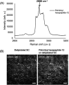
(A) The Raman spectrum of Palmitoyl hexapeptide-12 is in the high wavenumber region of the CH band. A prominent peak at 2886 cm–1 was selected for SRS imaging. (B) SRS images of a delipidated SC sample before and after treatment with the Palmitoyl hexapeptide-12 solution. Samples were dried before imaging. The morphology of the skin samples was visualized based on the SRS signals from the CH bonds. Scale bar: 20 μm.
Figure 4
Figure 4
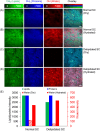
SRS imaging of SC samples. (A) Dry normal SC, (B) hydrated normal SC, (C) dry delipidated SC, and (D) hydrated delipidated SC. Green: SRS images at 2854 cm–1, attributed to the CH2 chemical bond vibration, represent total lipids. Blue: 2930 cm–1 for CH3 bonds, representing total proteins. Red: 3300 cm–1 for OH bonds, representing water distribution. (E) Averaged intensities of lipids, proteins, and water in the samples. Scale bar, 20 μm.
Figure 5
Figure 5
Time-lapse SRS imaging of the water content in SC samples enclosed in a chamber. The chamber was first purged with humidifying air flow for 40 min (flow rate: ∼1.0 L/min) and then purged with drying air flow for 50 min (flow rate: ∼0.5 L/min). (A) Normal SC, (B) delipidated SC, and (C) delipidated SC treated with the Palmitoyl hexapeptide-12 (0.45% w/v). Delipidated SC showed a much faster water loss under the drying flow through the chamber. (D) Application of Palmitoyl hexapeptide-12 treatment reduced water loss significantly on the delipidated SC samples. Green: lipids, blue: proteins, and red: water. Scale bar, 20 μm.
Figure 6
Figure 6
SC drying mechanics. Scatter plots of averaged SC contractile drying stress P_SC (A, B) and elastic modulus E_SC (C, D) measurements plotted against drying time for samples treated with DIW (yellow solid circle), 5% GLY (blue solid diamond), and Palmitoyl hexapeptide-12 (green solid triangle). Planes B and D do not include DIW treatment. Error bars denote a standard deviation of 3 ≤ n ≤ 5.
Figure 7
Figure 7
Final SC contractile drying stress, P_SC, and elastic modulus, E_SC. Bar graph of averaged final (t = 235 min) SC contractile drying stress P_SC (A) and elastic modulus E_SC (B) for samples treated with DIW, 5% GLY, and Palmitoyl hexapeptide-12. Error bars denote a standard deviation of 3 ≤ n ≤ 5. *, **, and ***, respectively note significant levels of p < 0.05, p < 0.01, and p < 0.001.
Figure 8
Figure 8
Co-assembly of palmitoyl hexapeptide-12 with human skin ECM components. (A) Gelling properties upon co-assembling of Palmitoyl hexapeptide-12 with hyaluronic acid (HAlow: 8–15 kDa, HAmid: 130–150 kDa and HAhigh: 750–1000 kDa), collagen type I and collagen-derived peptide ((GPP)2: GPPGPP and (GPP)3: GPPGPPGPP). (B) Rheological properties of co-assembled Palmitoyl hexapeptide-12-collagen type I gels. (C) Zeta potential, (D) Dynamic light scattering (DLS), and (E) circular dichroism analysis of Palmitoyl hexapeptide-12 co-assembling with collagen type I and HA molecules. (F) Scanning electron microscopy shows a characteristic diffusion-driven interface forming between co-assembling Palmitoyl hexapeptide-12 and collagen type I droplets. (G) Confocal microscopy of co-assembling Palmitoyl hexapeptide-12 and collagen type I solutions.
Figure 9
Figure 9
Representative H&E images and Van Gieson images of human skin before and after peptide treatment. (A) Ex vivo decellularized human skin treated with PBS as the control. (B) Ex vivo decellularized human skin treated with the Palmitoyl hexapeptide-12. Both images display a lack of blue/purple nuclear stain indicating effective decellularization. (C) Ex vivo decellularized human skin treated with PBS as the control. (D) Ex vivo decellularized human skin submerged in peptide solution showing a robust increase in elastin staining after 5 days of treatments. All images captured at 20× magnification. Scale bar: 50 μm.

References

    1. Cai H.; Wu F.-Y.; Wang Q.-L.; Xu P.; Mou F.-F.; Shao S.-J.; Luo Z.-R.; Zhu J.; Xuan S.-S.; Lu R.; Guo H.-D. Self-Assembling Peptide Modified with QHREDGS as a Novel Delivery System for Mesenchymal Stem Cell Transplantation after Myocardial Infarction. FASEB J. 2019, 33 (7), 8306–8320. 10.1096/fj.201801768RR. - DOI - PubMed
    1. Ligorio C.; Vijayaraghavan A.; Hoyland J. A.; Saiani A. Acidic and Basic Self-Assembling Peptide and Peptide-Graphene Oxide Hydrogels: Characterisation and Effect on Encapsulated Nucleus Pulposus Cells. Acta Biomater. 2022, 143, 145–158. 10.1016/j.actbio.2022.02.022. - DOI - PubMed
    1. Gelain F.; Luo Z.; Rioult M.; Zhang S. Self-Assembling Peptide Scaffolds in the Clinic. npj Regener. Med. 2021, 6 (1), 910.1038/s41536-020-00116-w. - DOI - PMC - PubMed
    1. Lindsey S.; Piatt J. H.; Worthington P.; Sönmez C.; Satheye S.; Schneider J. P.; Pochan D. J.; Langhans S. A. Beta Hairpin Peptide Hydrogels as an Injectable Solid Vehicle for Neurotrophic Growth Factor Delivery. Biomacromolecules 2015, 16 (9), 2672–2683. 10.1021/acs.biomac.5b00541. - DOI - PMC - PubMed
    1. Kassem S.; McPhee S. A.; Berisha N.; Ulijn R. V. Emergence of Cooperative Glucose-Binding Networks in Adaptive Peptide Systems. J. Am. Chem. Soc. 2023, 145 (17), 9800–9807. 10.1021/jacs.3c01620. - DOI - PubMed

LinkOut - more resources