. 2025 Feb 25;122(8):e2406715122.
doi: 10.1073/pnas.2406715122. Epub 2025 Feb 18.

A bacterial effector manipulates host lysosomal protease activity-dependent plasticity in cell death modalities to facilitate infection

Affiliations

A bacterial effector manipulates host lysosomal protease activity-dependent plasticity in cell death modalities to facilitate infection

Zhe Lu et al. Proc Natl Acad Sci U S A. .

Abstract

Crosstalk between cell death programs confers appropriate host anti-infection immune responses, but how pathogens co-opt host molecular switches of cell death pathways to reprogram cell death modalities for facilitating infection remains largely unexplored. Here, we identify mammalian cell entry 3C (Mce3C) as a pathogenic cell death regulator secreted by Mycobacterium tuberculosis (Mtb), which causes tuberculosis featured with lung inflammation and necrosis. Mce3C binds host cathepsin B (CTSB), a noncaspase protease acting as a lysosome-derived molecular determinant of cell death modalities, to inhibit its protease activity toward BH3-interacting domain death agonist (BID) and receptor-interacting protein kinase 1 (RIPK1), thereby preventing the production of proapoptotic truncated BID (tBID) while maintaining the abundance of pronecroptotic RIPK1. Disrupting the Mce3C-CTSB interaction promotes host apoptosis while suppressing necroptosis with attenuated Mtb survival and mitigated lung immunopathology in mice. Thus, pathogens manipulate host lysosomal protease activity-dependent plasticity in cell death modalities to promote infection and pathogenicity.

Keywords: Mycobacterium tuberculosis; RIPK1; cathepsin B; mammalian cell entry 3C; programmed cell death.


Conflict of interest statement

Competing interests statement:The authors declare no competing interest.

Figures

Fig. 1.
Fig. 1.
Identification of Mtb Mce3C as a cell death regulator that inhibits apoptosis and promotes necrosis of macrophages. (A) Schematic diagram of the procedure for identifying cell death regulators from the mce3 operon localizing in the RD7 of Mtb genome. (B) Representative histograms of fluorescence intensity of annexin V (Left) and PI (Right) in U937 cells. Cells were infected with BCG strains for 24 h. WT BCG infection group was used as control for multiple comparison test. (C) Confocal microscopy for caspase-3/7 activation (Left) or PI uptake (Right) in MDMs. Cells were infected with or without Mtb strains (green) for 24 h. Arrows indicate cells stained with active caspase-3/7 or PI (red). Nuclei (blue) were stained with DAPI. (Scale bar, 25 μm.) (D and E) Quantification of MDMs with caspase-3/7 activation (D) or with PI uptake (E). Cells were treated as in (C). Approximately 100 cells were examined. (F) Survival of Mtb in MDMs. Cells were infected with Mtb strains for 0 to 48 h. (G) In vitro growth kinetics of Mtb strains. (H) Bacterial loads of Mtb in the lungs of C57BL/6 mouse after infection for 0 to 28 d. (I) Histopathology of lung sections from C57BL/6 mice infected with Mtb strains for 0 to 28 d. (Scale bar, 100 μm.) (J) Quantitation of inflammatory areas in the lungs of mice treated as in (I). P > 0.05, ns; *P < 0.05; **P < 0.01; ***P < 0.001; ****P < 0.0001 [one-way ANOVA with Dunnett’s post hoc test for (B, D, and E) and two-way ANOVA with Tukey’s post hoc test for (F, H, and J)]. Data are shown as mean ± SEM. [n = 3 in (B); n = 4 in (DG); n = 5 mice per group in (HJ)]. Data are representative of at least three independent experiments.
Fig. 2.
Fig. 2.
Mtb Mce3C inhibits the apoptotic pathway while promoting the necroptotic pathway. (A) Immunoblotting analysis of the apoptotic pathway in U937 cells. Cells were infected with the indicated Mtb strains for 12 h in the presence or absence of 10 ng/mL TNF-α plus 10 μM CHX. DMSO served as a control. See also SI Appendix, Fig. S5A. (B) Immunoblotting analysis of the necroptotic pathway in U937 cells. Cells were infected with the indicated Mtb strains for 0 to 48 h. See also SI Appendix, Fig. S6A. Data are representative of at least three independent experiments.
Fig. 3.
Fig. 3.
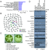
Mce3C reprograms host cell death pathways in a CTSB-dependent manner. (A and B) KEGG enrichment analysis (A) and the protein–protein interaction network (B) for host Mce3C-interacting proteins identified by yeast two-hybrid screening. A total of 73 mouse interacting proteins were identified. Proteins involved in the biological pathways related to lysosome, apoptosis, and necroptosis were colored with green, red, and blue, respectively. Lines indicate interactions between proteins inferred from STRING database. (C) Yeast two-hybrid assay for the interaction between Mce3C and mCTSB. Yeast strains were transformed with the indicated plasmids, where the interaction between TAK1 and TAB2 served as a positive control. Left, low stringency; Right, high stringency. (D) Immunoblotting for the cleavage of caspases, RIPK1, and PARP, the phosphorylation of RIPK1, RIPK3, and MLKL, and the mitochondrial release of cytochrome C (Cyto C) in U937 cells. Cells were infected with or without the indicated Mtb strains for 24 h.
Fig. 4.
Fig. 4.
CTSB cleaves BID and RIPK1 to promote apoptosis while inhibiting necroptosis. (A) Immunoblotting for the cleavage of caspase-8, BID, caspase-9, caspase-3, and PARP as well as the release of Cyto C from mitochondria (Mito.) into the cytosol (Cyto.) in U937 cells. Cells were treated with 10 ng/mL TNF-α plus 10 μM CHX in WT or CTSB-knockout (KO) cells for 12 h. (B) Immunoblotting for the cleavage of RIPK1 and caspase-8 and the phosphorylation of RIPK1, RIPK3, and MLKL in U937 cells. Cells were treated with 10 ng/mL TNF-α plus 10 µM Q-VD-Oph in WT or CTSB-KO cells for 12 h. (C) Schematic model for cleavage sites in RIPK1 by caspase-8 or CTSB. (D) Immunoblotting for the cleavage of RIPK1 or its mutants by caspase-8 or CTSB. Data are representative of at least three independent experiments.
Fig. 5.
Fig. 5.
Mce3C inhibits CTSB-mediated cleavage of BID and RIPK1, resulting in apoptosis inhibition and necroptosis promotion. (A) Schematic diagram of Mtb Mce3C and its truncated forms. (B) IP of the HC of CTSB by Mce3C or its MCE truncation from lysates of HEK293T cells. Cells were cotransfected with vectors encoding indicated proteins. (C and D) Immunoblotting for the CTSB-mediated cleavage of BID (C) and RIPK1 (D) after incubation with or without Mce3C or its ΔMCE mutant for 4 h. (E) Protease activity analysis of CTSB or its H278A mutant. (F) Immunoblotting for the indicated proteins in U937 cells. Cells were infected with or without the indicated Mtb strains for 24 h. (G) Survival of Mtb in WT or CTSB-KO U937 cells infected with Mtb strains for 0 to 48 h. (H) Immunohistochemistry of cleaved caspase-3 and p-MLKL in the lungs of C57BL/6 mice. Mice were infected with Mtb strains by aerosol (~100 CFUs) for 28 d. The boxed areas at the top are enlarged below. (Scale bar, 200 µm.) (I and J) Quantitation of cleaved caspase-3-positive (I) or p-MLKL-positive (J) cells in the lungs of mice treated as in (H). P > 0.05, ns; **P < 0.01; ***P < 0.001; ****P < 0.0001 [one-way ANOVA with Tukey’s post hoc test for (E, I, and J); two-way ANOVA with Tukey’s post hoc test for (G)]. Data are shown as mean ± SEM. [n = 4 in (E and G); n = 5 mice per group in (HJ)]. Data are representative of at least three independent experiments.
Fig. 6.
Fig. 6.
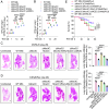
Disruption of the Mce3C–CTSB interaction attenuates Mtb survival and mitigates host inflammatory pathology. (A and B) Bacterial loads of Mtb in the lungs of C57BL/6 (A) or C3HeB/FeJ (B) mice. Mice were infected with Mtb strains by aerosol for 0 to 28 d. (C and D) Representative images and quantitation for inflammatory areas in the lungs of C57BL/6 (C) or C3HeB/FeJ (D) mice after infection for 28 d. (Scale bar, 100 μm.) (E) Survival curves of C57BL/6 and C3HeB/FeJ mice following Mtb infection. P > 0.05, ns; **P < 0.01; ***P < 0.001; ****P < 0.0001 [two-way ANOVA with Dunnett’s post hoc test for (A and B); one-way ANOVA with Dunnett’s post hoc test for (C and D); log rank test for (E)]. Data are shown as mean ± SEM. [n = 5 mice per group in (AD); n = 30 mice per group in (E)]. Data are representative of at least two independent experiments.

References

    1. Schaible U. E., et al. , Apoptosis facilitates antigen presentation to T lymphocytes through MHC-I and CD1 in tuberculosis. Nat. Med. 9, 1039–1046 (2003). - PubMed
    1. Jorgensen I., Rayamajhi M., Miao E. A., Programmed cell death as a defence against infection. Nat. Rev. Immunol. 17, 151–164 (2017). - PMC - PubMed
    1. Stutz M. D., et al. , Macrophage and neutrophil death programs differentially confer resistance to tuberculosis. Immunity 54, 1758–1771.e7 (2021). - PubMed
    1. Kim E. H., Wong S. W., Martinez J., Programmed necrosis and disease: We interrupt your regular programming to bring you necroinflammation. Cell Death Differ. 26, 25–40 (2019). - PMC - PubMed
    1. Snyder A. G., Oberst A., The antisocial network: Cross talk between cell death programs in host defense. Annu. Rev. Immunol. 39, 77–101 (2021). - PMC - PubMed

MeSH terms

Substances

LinkOut - more resources