. 2025 Feb 18;135(8):e187778.
doi: 10.1172/JCI187778. eCollection 2025 Apr 15.

Reduced EIF6 dosage attenuates TP53 activation in models of Shwachman-Diamond syndrome

Affiliations

Reduced EIF6 dosage attenuates TP53 activation in models of Shwachman-Diamond syndrome

Usua Oyarbide et al. J Clin Invest. .

Abstract

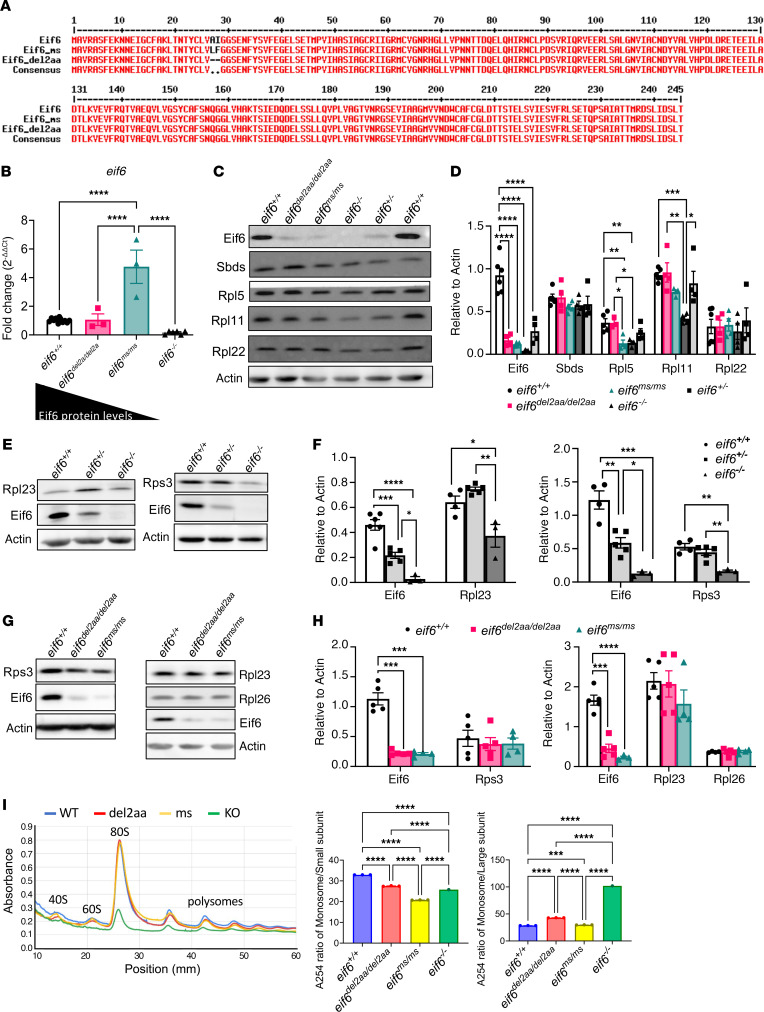
Shwachman-Diamond syndrome (SDS) is characterized by neutropenia, exocrine pancreatic insufficiency, and bony abnormalities with an increased risk of myeloid neoplasia. Almost all cases of SDS result from biallelic mutations in Shwachman-Bodian-Diamond syndrome (SBDS). SBDS interacts with elongation factor-like 1 (EFL1) to displace eukaryotic initiation factor 6 (EIF6) from the 60S ribosomal subunit. Released EIF6 permits the assembly of ribosomal large and small subunits in the cytoplasm. Decreased EIF6 levels due to haploinsufficiency or missense mutations, which lead to decreased protein expression, may provide a somatic genetic rescue and antileukemic effects. We observed accumulation of EIF6 protein in sbds-KO zebrafish models, confirmed this accumulation in patient-derived tissues, and correlated these with changes in ribosomal proteins and tumor protein p53 (TP53) pathways. The mechanism of action for this adaptive response is unknown. To address this, we generated eif6-KO zebrafish, which do not survive more than 10 days after fertilization. We also created 2 mutants with low Eif6 expression, i.e., 5%-25% of WT levels, that could survive until adulthood. We bred them with sbds-null strains and analyzed their phenotype and biochemical properties. Low Eif6 levels reduced Tp53 pathway activation but did not rescue neutropenia in Sbds-deficient zebrafish. Further studies elucidating the interplay between SBDS, EIF6, and TP53 and cellular stress responses offer promising insights into SDS pathogenesis, somatic genetic rescue, and therapeutic strategies.

Keywords: Bone marrow; Genetics; Hematology; Leukemias; Neutrophils; Oncology.


Conflict of interest statement

Conflict of interest: The authors have declared that no conflict of interest exists.

Figures

Figure 1
Figure 1. Eif6 accumulation in patients with SDS and sbds-KO zebrafish.
Western blots showing EIF6 accumulation in SDS lymphoblasts (A), SDS PBMCs (B), and sbds-KO fish from 5 dpf to 10 dpf (C). CTL1, healthy control no. 1. (D) IHC images of Eif6 expression showing accumulation in the liver and digestive tract (DT) of 10 dpf zebrafish. Original magnification, ×40.
Figure 2
Figure 2. Eif6 is essential for zebrafish embryonic development.
(A) Amino acid alignment of WT and eif6 KO zebrafish; the insertion of 1 bp causes early truncation. (B) mRNA eif6 levels by RT-qPCR on the tp53+/+ and tp53M214K/M214K backgrounds at 5 dpf. (C) Western blots showing the absence of Eif6 protein in the eif6–/– zebrafish larvae at 5 dpf. Note the dose effect in the eif6+/– fish compared with their WT siblings. (D) Western blot quantification. (E) eif6 KO showed a significantly lower number of neutrophils than the WT siblings at 5 dpf. (F) Sudan black staining for neutrophils counts (original magnification, ×2.5 and ×8). (G) Survival percentages for eif6+/+, eif6+/–, and eif6–/– siblings at 5, 8, and 10 dpf. *P < 0.05, **P < 0.01, and ***P < 0.001, by ANOVA. Data represent the mean ± SEM.
Figure 3
Figure 3. Low levels of Eif6 are enough for survival to adulthood, but only an absence of eif6 KO affects the levels of ribosomal proteins.
(A) Amino acid alignment of the 2 eif6 mutants (eif6ms/ms and eif6del2aa/del2aa) compared with the Eif6 WT sequence. (B) mRNA levels in all 3 eif6 mutants and WT zebrafish at 5 dpf. (C) Western blots showing Eif6, Sbds, and RP levels in the different eif6 mutants. (D) Western blot quantification. (E) Western blots showing Rpl23 and Rps3 in eif6+/+, eif6+/–, and eif6–/– siblings at 5 dpf. (F) Western blot quantification relative to actin. (G) Western blotting showing Rpl23 and Rps3 expression in eif6+/+, eif6del2aa/del2aa, and eif6ms/ms zebrafish at 5 dpf. (H) Western blot quantification relative to actin. (I) Polysome profiles of eif6 WT and mutants at 5 dpf. *P < 0.05, **P < 0.01, and ***P < 0.001, by 1-way ANOVA. Data represent the mean ± SEM.
Figure 4
Figure 4. Tp53 pathway activation in eif6 mutants does not affect survival rates.
RT-qPCR analysis of tp53 and its targets of WT and eif6 KO on the (A) tp53+/+ and (B) tp53M214K/M214K backgrounds. (C) Scheme of the double-heterozygote crosses (eif6+/– tp53+/M214K). (D) Percentage of survival for double-heterozygote crosses of eif6+/– tp53+/M214K at 8 and 15 dpf. (E) RT-qPCR analysis of tp53 and its targets (*P < 0.05, **P < 0.01, and ***P < 0.001, by 1-way ANOVA), and (F) UPR markers in WT and eif6ms/ms zebrafish at 5 dpf (*P < 0.05 and ***P < 0.001, by 2-tailed t test). Data represent the mean ± SEM.
Figure 5
Figure 5. Partial rescue of sbds KO with 1 copy of the WT eif6 allele.
(A) Scheme showing the incrossing of 2 double-heterozygotes (eif6+/– sbds+/–). (B) Genotype percentages at 15 dpf of an incrossing of double-heterozygotes. (C) Western blots showing Eif6 accumulation in sbds KO fish with only 1 copy of the eif6 WT allele (eif6+/–). (D) Western blot quantification of Eif6 protein expression. (E) Sudan black staining to detect the number of neutrophils in 10 dpf siblings. Scale bars: 200 μm and 50 μm. Data represent the mean ± SEM.
Figure 6
Figure 6. Correlation of Eif6 levels with tp53 activation in sbds-KO zebrafish.
(A) Scheme of sbds+/– crossings in the WT and Eif6 missense mutants and percentage of genotypes observed at 20 dpf. (B) Schemes of the 3 different crosses to obtain sbds WT and KO zebrafish on the eif6+/+, eif6+/ms, and eif6ms/ms backgrounds. (C) Immunoblot showing Eif6 accumulation in sbds-KO zebrafish. (D) Neutrophil counts at 10 dpf. *P < 0.05, **P < 0.01, ***P < 0.001, by 1-way ANOVA with Dunnett’s test. (E) Protein levels in sbds+/+, sbds+/–, and sbds–/–, in the eif6+/+ and eif6ms/ms mutant backgrounds, along with protein quantification. (F) RT-qPCR results for sbds and eif6 (G) and the tp53 pathway. *P < 0.05, **P < 0.01, ***P < 0.001, and ****P < 0.0001, by 1-way ANOVA with Tukey’s test. Data represent the mean ± SEM.
Figure 7
Figure 7. Tp53 activation in SDS patient–derived cells.
Western blots and quantification of (A) LCLs and (B) BM-MNCs. EIF6 mRNA (C) and protein (D) levels in 3 different siRNAs. (E) EIF6 mRNA levels in patients with SDS with and without siRNA (F) TP53 and CDKN1a mRNA levels are decreased after siRNA in patients with SDS. (G) Western blots showing decrease in TP53 levels after EIF6 knockdown using 3 different siRNAs. (H) TP53 protein quantification. Data represent the mean ± SEM. HD, healthy donor; NC, negative control.

References

    1. Rosenberg PS, et al. The incidence of leukemia and mortality from sepsis in patients with severe congenital neutropenia receiving long-term G-CSF therapy. Blood. 2006;107(12):4628–4635. doi: 10.1182/blood-2005-11-4370. - DOI - PMC - PubMed
    1. Boocock GR, et al. Mutations in SBDS are associated with Shwachman-Diamond syndrome. Nat Genet. 2003;33(1):97–101. doi: 10.1038/ng1062. - DOI - PubMed
    1. Zhang S, et al. Loss of the mouse ortholog of the shwachman-diamond syndrome gene (Sbds) results in early embryonic lethality. Mol Cell Biol. 2006;26(17):6656–6663. doi: 10.1128/MCB.00091-06. - DOI - PMC - PubMed
    1. Oyarbide U, et al. Loss of Sbds in zebrafish leads to neutropenia and pancreas and liver atrophy. JCI Insight. 2020;5(17):e134309. doi: 10.1172/jci.insight.134309. - DOI - PMC - PubMed
    1. Kawashima N, et al. Shwachman-Diamond syndromes: clinical, genetic, and biochemical insights from the rare variants. Haematologica. 2023;108(10):2594–2605. doi: 10.3324/haematol.2023.282949. - DOI - PMC - PubMed

MeSH terms