. 2025 Mar 5;147(9):7578-7587.
doi: 10.1021/jacs.4c16842. Epub 2025 Feb 18.

Fluorogenic Platform for Real-Time Imaging of Subcellular Payload Release in Antibody-Drug Conjugates

Affiliations

Fluorogenic Platform for Real-Time Imaging of Subcellular Payload Release in Antibody-Drug Conjugates

Ferran Nadal-Bufi et al. J Am Chem Soc. .

Abstract

Antibody-drug conjugates (ADCs) represent promising therapeutic constructs to enhance the selective delivery of drugs to target cells; however, attaining precise control over the timing and location of payload release remains challenging due to the complex intracellular processes that define ADC internalization, trafficking, and linker cleavage. In this study, we present novel real-time fluorogenic probes to monitor both subcellular dynamics of ADC trafficking and payload release. We optimized a tandem molecular design of sequential pH- and enzyme-activatable naphthalimide fluorophores to (1) track their subcellular localization along the endolysosomal pathway and (2) monitor linker cleavage with OFF-to-ON fluorescence switches. Live-cell imaging microscopy revealed that fluorogenic ADCs can traffic to the lysosomes and yet require residence time in these subcellular compartments for efficient linker cleavage. Notably, the compact size of fluorogenic naphthalimides did not impair the recognition of target cell surface reporters or the kinetics of payload release. This modular platform is applicable to many ADCs and holds promise to inform their rational design for optimal release profiles and therapeutic efficacy.


Conflict of interest statement

The authors declare the following competing financial interest(s): P.L.S., K.A.S, Z.W., R.D.W., V.L.M., and M.J.M. are employees of AbbVie. C.C.M. and A.D.H. were employees of AbbVie at the time of the study. AbbVie contributed in part to the design, study conduct, and financial support for this research.

Figures

Figure 1
Figure 1
Chemical design of fluorogenic probes for ADC imaging. The naphthalimide scaffold was modified with three orthogonal moieties: pH-sensitive amine groups to modulate fluorescence emission (green), cleavable linkers acting as switches for payload release (orange), and a succinimidyl ester group for direct conjugation to Lys residues in antibodies (gray).
Figure 2
Figure 2
Synthesis and characterization of a library of pH-sensitive naphthalimides. (a) General procedure for the synthesis of compounds A1-A26, including structures and synthetic yields of selected compounds A4, A10, A17, A18, A21, and A23. (b) Optical properties of compounds A1-A26, including excitation/emission maxima wavelengths, fluorescence fold increase ratios between pH 7.4 and pH 4.0, and pKa values. (c) Normalized fluorescence emission spectra of the 6 selected compounds (10 μM) in buffers ranging from pH 4.0 to pH 7.4 (λexc: 450 nm). Relative fluorescence quantum yields were calculated at pH 4.0 for neutral-to-acid compounds (A4, A10, A17, and A23) and at pH 7.4 for acid-to-neutral (A21) and always-on (A18) compounds.
Figure 3
Figure 3
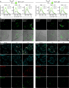
Fluorescent conjugates enable real-time subcellular imaging of antibody localization. (a) Normalized fluorescence emission spectra of the conjugates 8C11_A17 and 8C11_A21 (both at 200 nM) at pH 4.5 and pH 7.4 (λexc: 450 nm), and with varying degrees of labeling (FARs: ∼1.5, ∼3.0, and ∼8.0). For FAR calculations, the extinction coefficient of 8C11 was determined as 210,000 M–1 cm–1. (b) Representative fluorescence microscopy images of HEK293 cells transfected with mTNFα and incubated with 8C11_A17 (200 nM, left) and 8C11_A21 (200 nM, right) with different FARs after incubation for 2 h (exc/em: 450/550 nm). The presence of aggregates is highlighted by white arrows. The bottom row includes overlay images of fluorescence and brightfield. (c) Representative fluorescence microscopy images of live HEK293 cells transfected with mTNFα and treated with 8C11_A17 (200 nM, left) or 8C11_A21 (200 nM, right) at different time points (t: 30 min, 2 h, and 4 h). Subcellular activation of the fluorophores (A17 and A21) is highlighted by white arrows. Cells were costained with LysoTracker Red (50 nM, exc/em: 573/593 nm, red) and CellMask Deep Red (500 nM, exc/em: 660/675 nm, cyan). The bottom rows of the merged images include the single-channel fluorescence images of 8C11-dye conjugates, LysoTracker Red, and CellMask Deep Red. Scale bars: 20 μm.
Figure 4
Figure 4
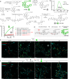
Design and synthesis of intramolecular quenchers to enable in situ monitoring of ADC linker cleavage. (a) Synthetic scheme for compounds A17-C and A17-C-AA. (b) Normalized fluorescence emission spectra of compounds A17, A17-C, and A17-C-AA at pH 4.0 (λexc: 450 nm). The fold decrease in maximal emission intensity between A17 and A17-C is highlighted in gray. (c) In vitro lysosomal payload release assays. A17-C or A17-C-AA (40 μM) were incubated with or without cathepsin B (40 nM) at pH 5.0 or 7.4, and with or without the cathepsin B inhibitor E64 (2 μM). Fluorescence fold increases were determined by referring to the caged compound A17-C. (d) Proposed mechanism for the tandem fluorescence activation of the dual-activated fluorophore 8C11_A17-C-AA. (e, f) Representative fluorescence microscopy images of live HEK293 cells transfected with mTNFα and treated with 200 nM 8C11_ A17 (e) or 8C11_A17-C-AA (f) (exc/em: 450/550) at different time points (t: 30 min, 2, 4, and 8 h). Cells were costained with LysoTracker Red (exc/em: 573/593 nm, red) and CellMask Deep Red (exc/em: 660/675 nm, cyan). Figure S13 contains the single-channel fluorescence images of 8C11-dye conjugates, LysoTracker Red and CellMask Deep Red. Scale bars: 20 μm.
Figure 5
Figure 5
Tracking linker cleavage and payload release in dual-conjugated ADCs. (a) Schematic representation of the mechanism of action of dual-conjugated anti-TNFα antibody (8C11) bearing both the payload GRM-103 (DAR: 4.0, represented by a gray star (inactive payload) or red star (active payload)) and the fluorophore (A17-C-AA, DAR: 3.0) attached via Ala-Ala linkers. (b) Luminescence-based cell reporter assay for measuring payload release. K562 cells, either transfected with a GRE reporter gene alone or cotransfected with mTNFα, were treated with serially diluted concentrations of ADCs (starting from 25 μg mL–1) for 72 h. Payload release was quantified by measuring firefly luciferase activity (RLU) after cell lysis. PBS and dexamethasone (100 nM) were used as negative and positive controls, respectively. Values presented as means and error bars as SEM (n = 3). (c) Representative fluorescence microscopy images of live HEK293 cells transfected with mTNFα treated with 200 nM 8C11_PL_A17-C-AA (exc/em: 450/550) at different time points (t: 30 min, 2, 4, and 8 h). Cells were costained with LysoTracker Red (exc/em: 573/593 nm, red) and CellMask Deep Red (exc/em: 660/675 nm, cyan). Scale bars: 20 μm.

References

    1. Dumontet C.; Reichert J. M.; Senter P. D.; Lambert J. M.; Beck A. Antibody-drug conjugates come of age in oncology. Nat. Rev. Drug Discovery 2023, 22, 641–661. 10.1038/s41573-023-00709-2. - DOI - PubMed
    1. Tsuchikama K.; Anami Y.; Ha S. Y. Y.; Yamazaki C. M. Exploring the next generation of antibody-drug conjugates. Nat. Rev. Clin. Oncol. 2024, 21, 203–223. 10.1038/s41571-023-00850-2. - DOI - PubMed
    1. Bross P. F.; Beitz J.; Chen G.; Chen X. H.; Duffy E.; Kieffer L.; Roy S.; Sridhara R.; Rahman A.; Williams G.; et al. Approval summary: gemtuzumab ozogamicin in relapsed acute myeloid leukemia. Clin. Cancer Res. 2001, 7, 1490–1496. - PubMed
    1. Ricart A. D. Antibody-drug conjugates of calicheamicin derivative: gemtuzumab ozogamicin and inotuzumab ozogamicin. Clin. Cancer Res. 2011, 17, 6417–6427. 10.1158/1078-0432.CCR-11-0486. - DOI - PubMed
    1. Mosele F.; Deluche E.; Lusque A.; Le Bescond L.; Filleron T.; Pradat Y.; Ducoulombier A.; Pistilli B.; Bachelot T.; Viret F.; et al. Trastuzumab deruxtecan in metastatic breast cancer with variable HER2 expression: the phase 2 DAISY trial. Nat. Med. 2023, 29, 2110–2120. 10.1038/s41591-023-02478-2. - DOI - PMC - PubMed

LinkOut - more resources