. 2025 Feb 18;15(1):5829.
doi: 10.1038/s41598-025-88380-y.

42 induces stress granule formation via PACT/PKR pathway

Affiliations

42 induces stress granule formation via PACT/PKR pathway

Vijay Sankar Ramasamy et al. Sci Rep. .

Abstract

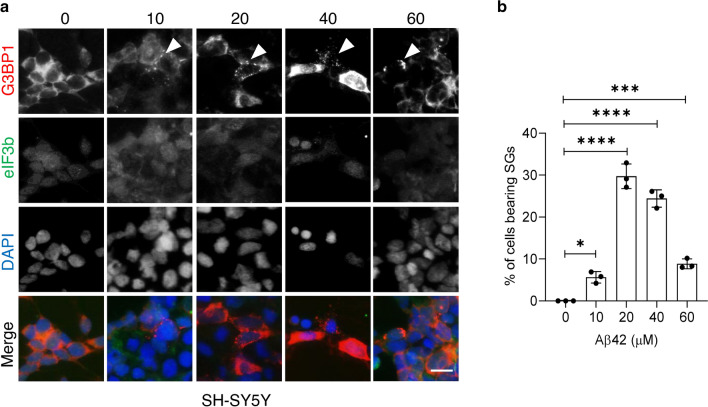
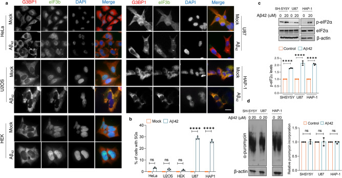
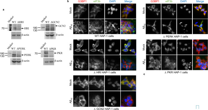
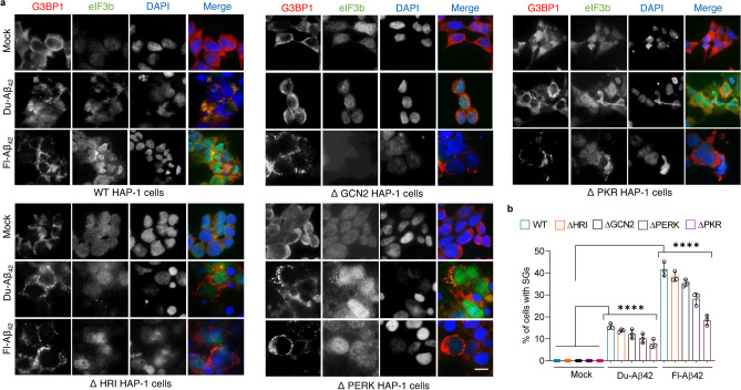
Stress granule (SG) formation has been linked to several neurodegenerative disorders, such as Alzheimer's disease (AD). Amyloid-β42 (Aβ42) is a key player in the pathogenesis of AD and is known to trigger various stress-related signaling pathways. However, the impact of Aβ on SG formation has not been fully understood. The primary aim of this study is to analyze the SG-inducing properties of Aβ42 and to uncover the molecular mechanisms underlying this process. Our results revealed that exposure to 20 μM Aβ42 led to a significant SG formation in neuroblastoma-derived (SH-SY5Y) and glioma-derived (U87) cell lines. Interestingly, we observed elevated levels of p-eIF2α, while overall protein translation levels remained unchanged. Monomeric and oligomeric forms of Aβ42 exhibited a 4-5 times stronger ability to induce SG formation compared to fibrillar forms. Additionally, treatment with familial mutants of Aβ42 (Dutch and Flemish) showed distinct effects on SG induction. Moreover, our findings using eIF2α kinases knockout (KO) cell lines demonstrated that Aβ-induced SG formation is primarily dependent on Protein Kinase R (PKR). Subsequent proximity ligation assay (PLA) analysis revealed a close proximity of PACT and PKR in Aβ-treated cells and in AD mouse hippocampus. Taken together, our study suggests that Aβ42 promotes SG formation through PKR kinase activation, which in turn requires PACT involvement.

Keywords: Alzheimer’s; Amyloid-β; Aβ42; PKR kinase; Stress granule; Stress signaling.


Conflict of interest statement

Declarations. Competing interests: The authors declare no competing interests.

Figures

Fig. 1
Fig. 1
Aβ42 exposure induced SG formation in SH-SY5Y cells. (a) cells were treated with indicated concentrations of Aβ42 for 24 h and then immunostained with SG markers eIF3b (green), G3BP1 (red), and nuclei stain DAPI (blue). Scale bar, 10 μm. (b) SG quantification data showing the percentage of cells harboring SGs. one-way analysis of variance (ANOVA) at the 95% confidence interval followed by a Dunnett’s post-hoc test. Error bars indicate standard deviation (S.D.) (n = 3). **p < 0.01, ***p < 0.001, ****p < 0.0001.
Fig. 2
Fig. 2
Aβ42 exposure increased p-eIF2α levels but has no significant effect on global protein translation. (a) HeLa, U2OS, HEK, U87, and HAP-1 cell lines were seeded on coverslips with appropriate media in 4- well plates, grown and treated with 20 μM Aβ42 for 24 h. After that, cells were immunostained with eIF3b (green), G3BP1 (red) and nuclei stain DAPI (blue). Scale bar, 10 μm. (b) Bar graph shows the percentage of cells containing SGs. (one-way ANOVA at the 95% confidence interval followed by Bonferroni’s post-hoc test, ****P < 0.001) Error bars indicate S.D. (n = 3), (c) Representative image of immunoblots and quantitative analysis showing the expression levels of p-eIF2α and eIF2α in Aβ42 treated cell lines. U87, SH-SY5Y, and HAP-1 cells were exposed to 20 μM Aβ42 for 24 h and the whole cell lysates were subjected to immunoblot. (one-way ANOVA, ****p < 0.0001) (d) Representative image of immunoblots and quantitative analysis showing the levels of puromycylated proteins under Aβ42 treatment. U87, SH-SY5Y, and HAP-1 cells were exposed to 20 μM Aβ42 followed by 10 min puromycin pulse (10 mg/ml) and the whole cell lysates were subjected to immunoblot (one-way ANOVA, ns).
Fig. 3
Fig. 3
Monomeric and oligomeric Aβ42 display better SG- inducing characteristics. (a) Representative images of G3BP1 and eIF3b immunofluorescence staining of SH-SY5Y cells treated with different conformers of Aβ42. Scale bar, 10 μm. (b) Bar graph shows the percentage of cells containing SGs. one-way ANOVA at the 95% confidence interval followed by Dunnett’s post-hoc test, ****p < 0.0001. Error bars indicate S.D. (n = 3), (c) Representative image of immunoblots showing amount of p-eIF2α in Aβ42 treated SH-SY5Y cells. (d) Representative images of G3BP1 and eIF3b immunofluorescence staining of U87 cells treated with different conformers of Aβ42. Scale bar, 10 μm. (e) Bar graph shows the percentage of cells containing SGs. one-way ANOVA at the 95% confidence interval followed by Dunnett’s post-hoc test, **p < 0.01, ***p < 0.001, ****p < 0.0001. Error bars indicates S.D. (n = 3) (f) Representative image of immunoblots showing expression level of p-eIF2α and eIF2α in Aβ42 treated U87 cells.
Fig. 4
Fig. 4
Dutch- and Flemish-Aβ42 display distinct SG- inducing characteristic. (a) Representative images of G3BP1 and eIF3b immunofluorescence staining of SH-SY5Y cells treated with Du- and Fl-Aβ42 for 24 h. Scale bar, 10 μm. (b) Bar graph shows the percentage of cells containing SGs. one-way ANOVA at the 95% confidence interval followed by Dunnett’s post-hoc test, ****p < 0.0001. Error bars indicate S.D. (n = 3), (c) Representative image of immunoblots showing amount of p-eIF2α and eIF2α in Du- and Fl-Aβ42 treated SH-SY5Y cells. (d) Representative images of G3BP1 and eIF3b immunofluorescence staining of U87 cells treated with Du- and Fl-Aβ42. (e) Bar graph shows the percentage of cells containing SGs. one-way ANOVA at the 95% confidence interval with Dunnett’s post-hoc test, ***p < 0.001, ****p < 0.0001. Error bars indicate S.D. (n = 3). (f) Representative image of immunoblots showing amount of p-eIF2α in Du- and Fl-Aβ42 treated U87 cells.
Fig. 5
Fig. 5
Aβ42 induced SG formation is PKR-dependent. (a) Immunoblot analysis of HRI, GCN2, PERK, and PKR protein expression levels in HAP-1 WT and KO cell lines. Whole cell lysates of HAP-1 WT and KO cell lines were subjected to immunoblot with respective antibodies (b) Representative images of G3BP1 and eIF3b immunofluorescence staining of HAP-1-WT and -KO cells treated with Aβ42. Scale bar, 10 μm. (c) Bar graph shows the percentage of cells containing SGs. one-way ANOVA with Bonferroni’s post-hoc test, *p < 0.05, ****p < 0.0001. Error bars indicate S.D. (n = 3).
Fig. 6
Fig. 6
Du- and Fl-Aβ42 induced SG formation is PKR-dependent. (a) Representative images of G3BP1 and eIF3b immunofluorescence staining of HAP-1-WT and -KO cells treated with Du- and Fl-Aβ42 for 24 h. Scale bar, 10 μm. (b) Bar graph shows the percentage of cells containing SGs. one-way ANOVA at the 95% confidence interval with Bonferroni’s post-hoc test, ****p < 0.0001. Error bars indicate S.D. (n = 3).
Fig. 7
Fig. 7
Aβ42 induced SG formation requires PACT: PKR interaction. (a) Representative images of PACT/PKR (red) proximity ligation assay of SH-SY5Y and U87 cells treated with Aβ42. Nuclei stained with DAPI (blue) Scale bar, 10 μm. (b) Bar graph shows the percentage of PACT/PKR positive cells. Two-tailed T-test, ****P < 0.0001 Error bars indicate S.D. (n = 2), (c) Representative Images of PACT/PKR (red) proximity ligation assay of wild type and 5XFAD mouse hippocampus. Nuclei stained with DAPI (blue). Scale bar, 50 μm.

References

    1. Palangi, F., Samuel, S. M., Thompson, I. R., Triggle, C. R. & Emara, M. M. Effects of oxidative and thermal stresses on stress granule formation in human induced pluripotent stem cells. PLoS ONE12, e0182059. 10.1371/journal.pone.0182059 (2017). - PMC - PubMed
    1. Sun, C. L., Van Gilst, M. & Crowder, C. M. Hypoxia-induced mitochondrial stress granules. Cell Death Dis.14, 448. 10.1038/s41419-023-05988-6 (2023). - PMC - PubMed
    1. Iadevaia, V. et al. Novel stress granule-like structures are induced via a paracrine mechanism during viral infection. J. Cell Sci.10.1242/jcs.259194 (2022). - PMC - PubMed
    1. Protter, D. S. W. & Parker, R. Principles and properties of stress granules. Trends Cell Biol.26, 668–679. 10.1016/j.tcb.2016.05.004 (2016). - PMC - PubMed
    1. Gilks, N. et al. Stress granule assembly is mediated by prion-like aggregation of TIA-1. Mol. Biol. Cell15, 5383–5398. 10.1091/mbc.e04-08-0715 (2004). - PMC - PubMed

MeSH terms

LinkOut - more resources