Review
. 2025 Mar;44(10):637-651.
doi: 10.1038/s41388-025-03300-8. Epub 2025 Feb 18.

Deletion of 17p in cancers: Guilt by (p53) association

Affiliations
Review

Deletion of 17p in cancers: Guilt by (p53) association

Francisca van Kampen et al. Oncogene. 2025 Mar.

Abstract

Monoallelic deletion of the short arm of chromosome 17 (del17p) is a recurrent abnormality in cancers with poor outcomes. Best studied in relation to haematological malignancies, associated functional outcomes are attributed mainly to loss and/or dysfunction of TP53, which is located at 17p13.1, but the wider impact of deletion of other genes located on 17p is poorly understood. 17p is one of the most gene-dense regions of the genome and includes tumour suppressor genes additional to TP53, genes essential for cell survival and proliferation, as well as small and long non-coding RNAs. In this review we utilise a data-driven approach to demarcate the extent of 17p deletion in multiple cancers and identify a common loss-of-function gene signature. We discuss how the resultant loss of heterozygosity (LOH) and haploinsufficiency may influence cell behaviour but also identify vulnerabilities that can potentially be exploited therapeutically. Finally, we highlight how emerging animal and isogenic cell line models of del17p can provide critical biological insights for cancer cell behaviour.


Conflict of interest statement

Competing interests: The authors declare no competing interests financial or otherwise Preparation of this article was supported by a Northwest Cancer Research UK and Bloom Appeal (NWCRTBA2021.02) grant funding to AC, MG, ARP, NK and JRS.

Figures
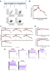
Fig. 1
Fig. 1. Analysis of deletion size associated with 17p aneuploidy in various cancers.
A Dot plots showing copy number alteration (CNA) data of the four indicated genes (upper panel shows where they are located on chromosome 17p) plotted relative to TP53 CNA. The CNA data is derived from the cancer cell lines available on DepMap. B Graph showing the frequency of genes located on 17p that are co-deleted with TP53 in a continuous fashion, giving insight into the size of deletion that is most commonly observed in cells bearing del17p. The location of TP53 is indicated by the dotted red line, as are the positions of the genes illustrated in part A. The data used to construct this graph is taken from all cell lines available on DepMap. C Graphs showing the frequency of genes located on 17p that are co-deleted with TP53 in a continuous fashion using data (from DepMap) associated with cell lines derived from the indicated cancers. The location of TP53 is indicated by the dotted red line, as are the positions of the genes illustrated in part A. D Graphs showing the frequency of genes located on 17p that are co-deleted with TP53 in a continuous fashion using data (from cBioPortal) associated with primary cells derived from the indicated cancers. The location of TP53 is indicated by the dotted red line, as are the positions of the genes illustrated in part A. E Hierarchical cluster plots indicating 17p deletion size (x-axis) within cells from individual patients (y-axis) calculated from data recorded in cBioPortal for primary cells from the indicated cancers.
Fig. 2
Fig. 2. Analysis of 17p deletion in relation to mutation of TP53.
Data from cBioPortal on mutation within TP53 was analysed against the presence or absence of del17p for the indicated cancers (brca breast invasive carcinoma, coad colorectal adenocarcinoma, dlbc diffuse large B cell lymphoma, laml acute myeloid leukaemia, paad pancreatic adenocarcinoma, prad prostate adenocarcinoma) and illustrated according to A count within the total data set available, or as B a percentage within each variable (loss vs. intact 17p). TP53 mutation classification is indicated by the colour scheme.
Fig. 3
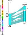
Fig. 3. Human chromosome 17 is syntenic with mouse chromosome 11B.
A chromosome map comparing gene arrangement between mouse chromosome 11B and human chromosome 17. The cytobands 11B2, 11B3 and 11B4 are marked for reference to human 17p.
Fig. 4
Fig. 4. Upset analysis of gene expression in primary malignant cells with del17p.
Gene expression in primary malignant cells with or without del17p was compared (with DESeq2), focusing on genes located on 17p that were downregulated in del17p cells. Set size refers to the number of genes showing change in cells from the indicated cancers. Intersection size refers to the shared genes between the different cancers analysed. Cells from 5 cancers were compared; laml acute myeloid leukaemia, prad prostate adenocarcinoma, paad pancreatic adenocarcinoma, brca breast invasive carcinoma, coad colorectal adenocarcinoma. The data for analysis was taken from cBioPortal.
Fig. 5
Fig. 5. Enrichment pathway analysis of genes on 17p affected by LOH and potential haploinsufficiency.
The panel of 76 genes identified from the UpSet plot analysis was subjected to enrichment pathway analysis using the Metascape tool (https://metascape.org). Reactome and GO terms are listed according to their probability score.
Fig. 6
Fig. 6. Positioning and REACTOME analysis of microRNA coding sequences located on 17p.
A Pie chart showing the frequency of microRNA genes located within their own host gene, or within promoter or enhancer regions / intronic gene sequences of other genes. B Cytoscape EnrichmentMap visualisation of DAVID REACTOME pathway enrichment analysis of predicted targets with a target score of >90 for all 17p-derived miRNAs. The sizes of each node represent the number of genes within that particular pathway, while the node colour indicates the False Discovery Rate q significance value. The thickness of each connection indicates the number of genes shared between adjacent nodes (p < 0.01, q < 0.1, overlap threshold = 0.5).
Fig. 7
Fig. 7. Role of del17p in tumorigenesis and therapy resistance/sensitivity.
A Summary of the role del17p plays in tumorigenesis. del17p is an early event where selection processes dictate expansion of the clone which, in turn, adds to therapy resistance and increased aggressive behaviour / metastatic potential. The inset list indicates the cancer types where del17p is associated with aggressive tumour behaviour and poor clinical outcome. CLL chronic lymphocytic leukaemia, MM multiple myeloma, AML acute myeloid leukaemia, DLBCL diffuse large B cell lymphoma, MCL mantle cell lymphoma, ALL acute lymphoblastic leukaemia, HCC hepatocellular carcinoma, CRC colorectal carcinoma. B Summary of the impact of del17p on malignant cell vulnerability and resistance to therapy. Vulnerability / Therapy resistance likely results from LOH and haploinsufficiency of the listed cell-essential genes or TSGs, respectively.

References

    1. Murphy SJ, Hart SN, Halling GC, Johnson SH, Smadbeck JB, Drucker T, et al. Integrated genomic analysis of pancreatic ductal adenocarcinomas reveals genomic rearrangement events as significant drivers of disease. Cancer Res. 2016;76:749–61. - PMC - PubMed
    1. Liu Y, Chen C, Xu Z, Scuoppo C, Rillahan CD, Gao J, et al. Deletions linked to TP53 loss drive cancer through p53-independent mechanisms. Nature. 2016;531:471–5. - PMC - PubMed
    1. Ben-David U, Amon A. Context is everything: aneuploidy in cancer. Nat Rev Genet. 2020;21:44–62. - PubMed
    1. Li Y, Sun Y, Kulke M, Hechler T, Van der Jeught K, Dong T, et al. Targeted immunotherapy for HER2-low breast cancer with 17p loss. Sci Transl Med. 2021;13:eabc6894. - PMC - PubMed
    1. Harada T, Chelala C, Bhakta V, Chaplin T, Caulee K, Baril P, et al. Genome-wide DNA copy number analysis in pancreatic cancer using high-density single nucleotide polymorphism arrays. Oncogene. 2008;27:1951–60. - PMC - PubMed

Substances

Supplementary concepts